
Regenerating functional neurons for treatment of hemorrhagic stroke

Pending Publication Date: 2021-06-03
PENN STATE RES FOUND
View PDF11 Cites 0 Cited by
  • Summary
  • Abstract
  • Description
  • Claims
  • Application Information

AI Technical Summary

Benefits of technology

This patent describes a method for generating new brain cells, increasing the survival of certain brain cells, and reducing the number of reactive astrocytes in a mammal that has had a hemorrhagic stroke. The method involves administering a composition containing specific proteins to the mammal. The composition can be delivered using an expression vector or a recombinant viral or adeno-associated virus expression vector. The method can help to improve the recovery and outcomes of mammals that have had a hemorrhagic stroke.

Problems solved by technology

When that happens, part of the brain cannot get the blood (and oxygen) it needs, so it and brain cells die.
However, there remains a significant unmet need for treatment of patients having had a hemorrhagic stroke.

Method used


Image

  • Regenerating functional neurons for treatment of hemorrhagic stroke
  • Regenerating functional neurons for treatment of hemorrhagic stroke
  • Regenerating functional neurons for treatment of hemorrhagic stroke

Examples


example 1

of Intracerebral Hemorrhage

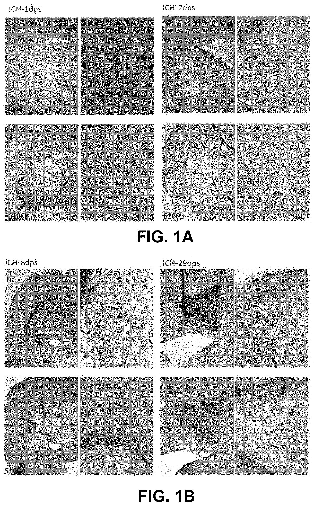
[0151]0.2 μL collagenase was injected to mouse striatum. After 1 day, 2 days, 8 days, and 29 days, data were collected, and DAB and iron staining were conducted. FIG. 1A-1B showed the Iba1 and S100b DAB staining with iron staining from 1 day to 29 days post induction of ICH.

[0152]These results showed the morphological changes of astrocytes and microglia after ICH as well as the process of accumulation of ferric iron. These results provided a reference to choose time points to intervene to treat ICH.

example 2

onversion of Reactive Astrocytes to Neurons in a Mouse Model of Intracerebral Hemorrhage (Short Term)

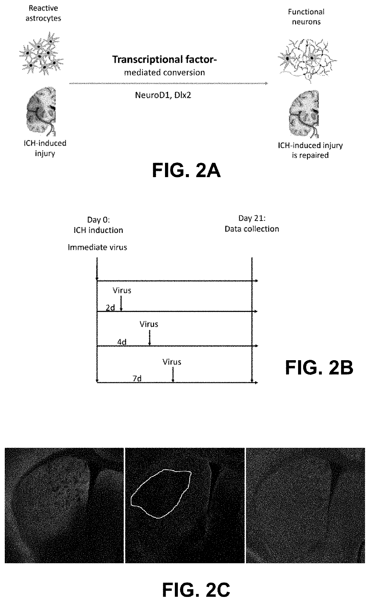
[0153]A set of experiments was performed to assess the in vivo conversion of reactive astrocytes into neurons following treatment with AAV5 viruses encoding NeuroD1 and Dlx2. ICH induction at day 0 was performed by injecting 0.2 μL of collagenase into striatum. Mice were injected with 1 μL of AAV5-GFA104-cre: 3×10¹¹, 1 μL of AAV5-CAG-flex-GFP: 3.4×10¹¹, 1 μL of AAV5-CAG-flex-ND1-GFP: 4.55×10¹¹, or 1 μL of AAV5-CAG-flex-Dlx2-GFP: 2.36×10¹² at 2 days, 4 days, and 7 days post ICH induction. On day 21, data regarding astrocyte conversion were collected.

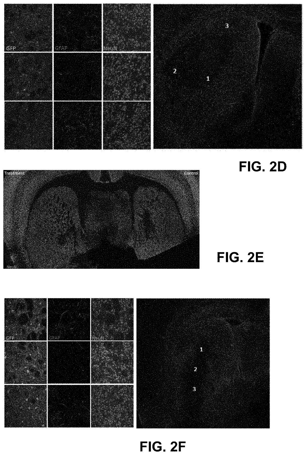
[0154]FIG. 2A-2B showed the schematics of the experiments about in vivo conversion in short term. Different virus injection times (immediately, 2 dps, 4 dps, and 7 dps) were conducted to find the optimal time window to repair ICH. FIG. 2C-2P revealed the immunostaining of GFP, GFAP, and NeuN, accordingly. The results consistently showed th...

example 3

onversion of Reactive Astrocytes to Neurons in a Mouse Model of Intracerebral Hemorrhage (Long Term)

[0156]A set of experiments was performed to assess the in vivo conversion of reactive astrocytes into neurons following treatment with AAV5 viruses encoding NeuroD1 and Dlx2. ICH induction at day 0 was performed by injecting 0.35 μL of collagenase into striatum. Mice were injected with 1 μL of AAV5-GFA104-cre: 3×10¹¹, 1 μL of AAV5-CAG-flex-GFP: 3.4×10¹¹, 1 μL of AAV5-CAG-flex-ND1-GFP: 4.55×10¹¹, or 1 μL of AAV5-CAG-flex-Dlx2-GFP: 2.36×10¹² at 2 days and 7 days post ICH induction. Two months post induction, mice were harvested, and data were collected.

[0157]FIG. 3A shows the experimental design of the long-term repair effect of ND1 and Dlx2 on ICH. FIG. 3B-3G present the immunostaining of GFP, GFAP, and NeuN. FIG. 3B-3C showed almost all the GFP-positive cells had neuronal morphologies and expressed NeuN two months after virus infection when the virus was injected immediately after ICH...


PUM

PropertyMeasurementUnit
Timeaaaaaaaaaa
Volumeaaaaaaaaaa
Volumeaaaaaaaaaa

Abstract

This document provides methods and materials involved in treating mammals having had a hemorrhagic stroke. For example, methods and materials for administering a composition containing exogenous nucleic acid encoding a NeuroD1 polypeptide and exogenous nucleic acid encoding a Dlx2 polypeptide to a mammal having had a hemorrhagic stroke are provided.

Description

CROSS-REFERENCE TO RELATED APPLICATIONS[0001]This application claims benefit to U.S. Application Ser. No. 62 / 916,706, filed on Oct. 17, 2020, the contents of this aforementioned application being fully incorporated here by reference.BACKGROUND1. Technical Field[0002]This document relates to methods and materials involved in treating mammals having had a hemorrhagic stroke. For example, this document provides methods and materials for administering a composition containing exogenous nucleic acid encoding a NeuroD1 polypeptide (or a biologically active fragment thereof) and nucleic acid encoding a Dlx2 polypeptide (or a biologically active fragment thereof) to a mammal having had a hemorrhagic stroke.2. Background Information[0003]Stroke is a disease that affects the arteries leading to and within the brain. It is the number five cause of death and a leading cause of disability in the United States. A stroke occurs when a blood vessel that carries oxygen and nutrients to the brain is ...

Claims


Application Information

Patent Timeline
IPC IPC(8): A61K38/17A61K9/00A61P25/00
CPCA61K38/1703A61K48/00A61P25/00A61K9/0085C07K14/47C07K14/4702C12N5/0619A61K48/005A61K48/0075C12N2750/14143
Inventor CHEN, GONG
Owner PENN STATE RES FOUND
Learn More