Fluorescence imaging system

Pending Publication Date: 2021-02-25
REBOUND THERAPEUTICS CORP
View PDF1 Cites 2 Cited by
  • Summary
  • Abstract
  • Description
  • Claims
  • Application Information

AI Technical Summary

Benefits of technology

The patent describes a device and method that can help surgeons see diseased tissue during minimally invasive surgery. The device includes a camera that can capture images of the tissue using a broad spectrum light, which can illuminate the tissue. Additionally, there is an excitation light source that can cause the tissue to fluoresce, which can be used to visualize any disease-related tissue. The camera alternately shows the white light images and the blue light images, which can help the surgeon see both the target tissue and any fluorescing tissue simultaneously on the same screen. This improves the surgeon's ability to visualize and excise the diseased tissue while avoiding damage to healthy tissue. The method involves placing the camera and lights in close proximity to the surgery and controlling the camera to obtain images of the target tissue and diseased tissue. The control system alternately shows the broad spectrum light images and the narrow spectrum excitation light images, which are generated in rapid alternating fashion. The method allows for real-time visualization of diseased tissue without the need for switching between views or interrupting the surgery.

Problems solved by technology

Especially in the brain, a surgeon wants to remove cancerous tissue while avoiding disruption of healthy tissue, but the difficulty in discerning one from the other makes this difficult.
However, during narrow spectrum illumination, the surgical workspace is dark, being illuminated with only the excitation light, so that surrounding healthy tissue cannot be clearly seen.

Method used


Image

  • Fluorescence imaging system
  • Fluorescence imaging system
  • Fluorescence imaging system

Examples


Embodiment Construction

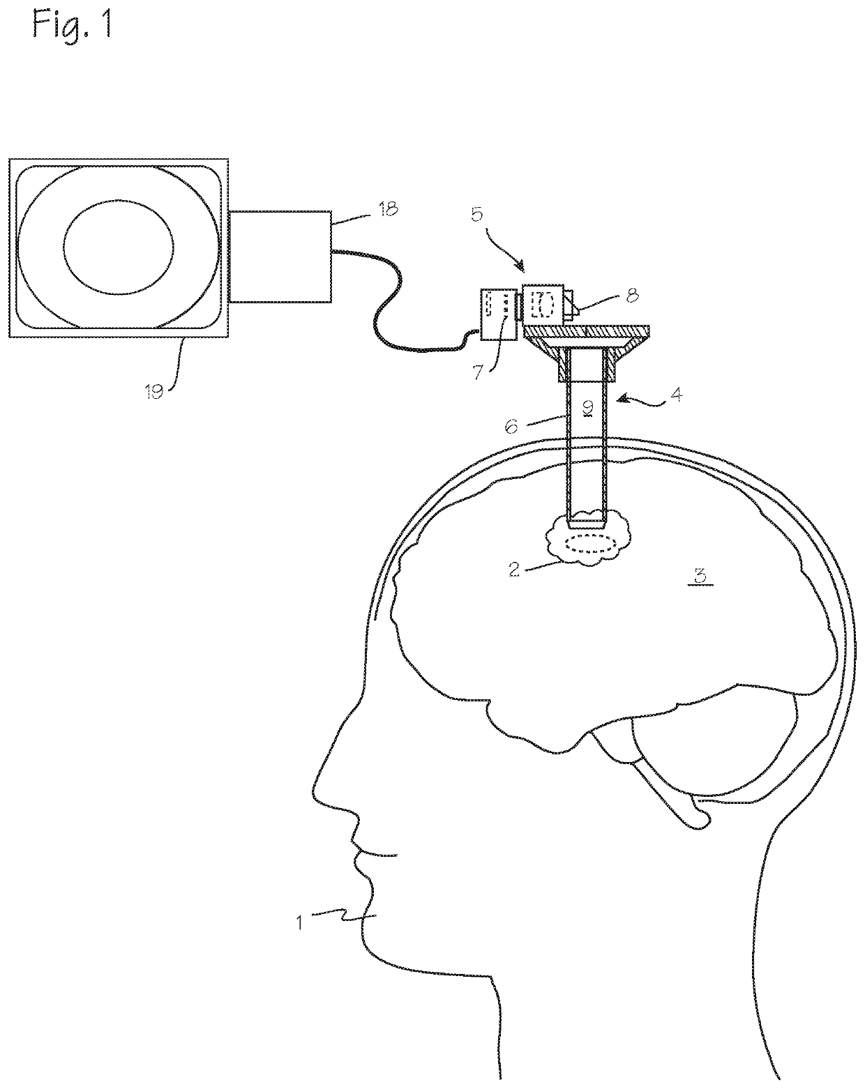
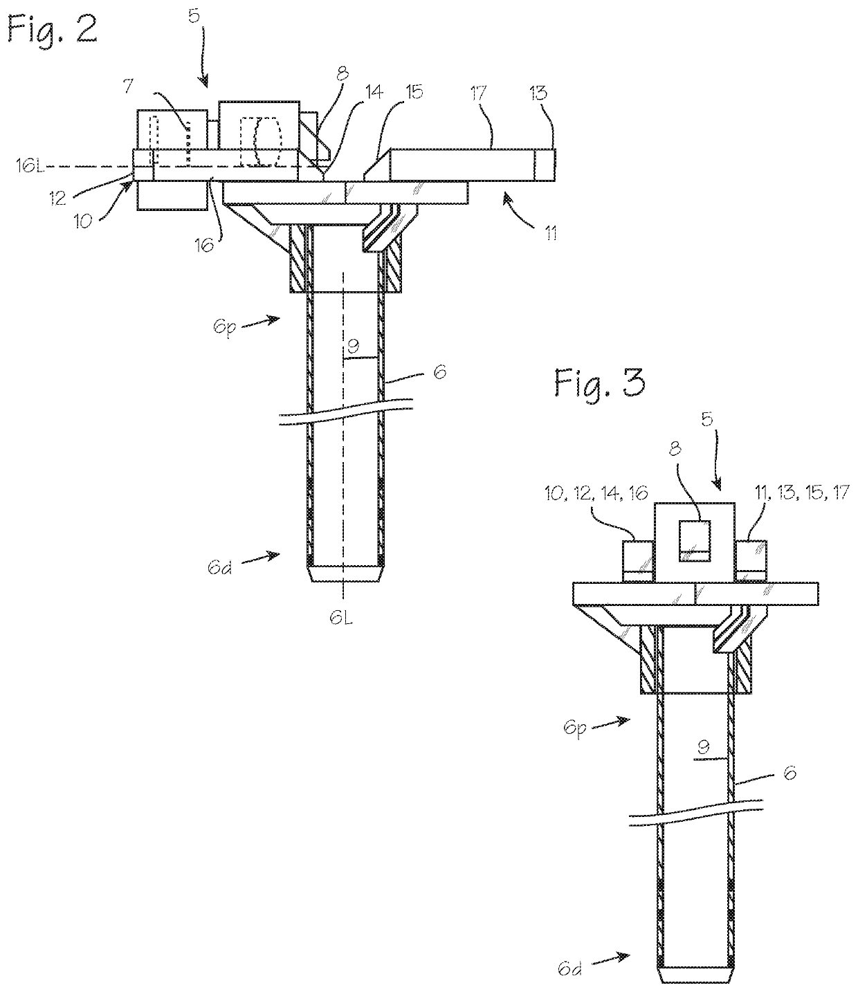
[0015]FIGS. 1, 2 and 3 illustrate a cannula system that may be conveniently used to implement the imaging method describe in relation to FIGS. 4 through 8 in a minimally invasive surgery. FIG. 1 illustrates a patient 1 with diseased tissue 2 in the brain 3 that necessitates surgical intervention, with a cannula 4 which has been inserted into diseased tissue, with the distal end of the cannula proximate the diseased tissue. The diseased tissue may be a glioma or glioblastoma in the brain, an ependymoma in the spine, or other diseased tissue. A camera 5 is mounted on the proximal rim of the cannula, with a portion of the camera overhanging the rim of the cannula and disposed over the lumen of the cannula, and is operable to obtain video or still images of the distal end of the cannula lumen, including target tissue at the distal end of the cannula such as the brain and any diseased tissue in the brain. As shown in both FIGS. 1, 2 and 3, the cannula comprises a cannula tube 6 with the ...


PUM


Abstract

A system for fluorescence guided surgery which includes alternatingly obtaining white light images and blue light images and simultaneously displaying those images on a display screen, such that a surgeon is presented with immediate, real-time video which shows the target tissue under white light and fluorescing tissue under blue light on the same screen, at the same time. The images may be presented side-by-side, or superimposed.

Description

FIELD OF THE INVENTIONS[0001]The inventions described below relate to the field of minimally invasive surgery for the treatment of tumors.BACKGROUND OF THE INVENTIONS[0002]Fluorescence Guided Surgery is a technique used to identify cancerous tumors and cancerous cells during surgery. Under broad spectrum light (white light), there is no clear difference in the appearance of cancerous tumor and cancerous cells and surrounding healthy tissue, especially at the margins of the cancerous tumor. Especially in the brain, a surgeon wants to remove cancerous tissue while avoiding disruption of healthy tissue, but the difficulty in discerning one from the other makes this difficult. Improved visualization of the cancer can help ensure that all cancerous tissue has been removed while reducing damage to healthy tissue such as nerves, blood vessels, and brain tissue.[0003]To use the Fluorescence Guided Surgery technique, a fluorescent agent is administered to the patient. Shortly after administr...

Claims


Application Information

Patent Timeline
IPC IPC(8): A61B5/00A61K49/00A61B17/34
CPCA61B5/0071A61B5/7425A61B5/0077A61B2017/3425A61K49/0034A61B17/3423A61K49/0036A61B2090/103A61B17/3421A61B90/37A61B2090/373A61B5/6865A61B5/0082A61B5/0036A61B5/0042
Inventor TSUKASHIMA, ROSSAMORSOLO, MILO ALFARO
Owner REBOUND THERAPEUTICS CORP
Why Eureka
Try Eureka