Closed-loop system for contextually-aware image-quality collection and feedback

a closed-loop system and image-quality collection technology, applied in the field of medical arts, medical acquisition arts, medical reporting arts, can solve the problems of interpreter not being able to draw diagnostic conclusions, not generally qualified to perform medical diagnosis, and sometimes acquiring images that are not optimal or even non-diagnostic, so as to improve the user interface

Pending Publication Date: 2019-08-29
KONINKLJIJKE PHILIPS NV
View PDF3 Cites 10 Cited by
  • Summary
  • Abstract
  • Description
  • Claims
  • Application Information

AI Technical Summary

Benefits of technology

The patent text talks about an improved medical workstation with a better user interface. The technical effect of this is that it provides a more efficient and effective tool for medical professionals to work with.

Problems solved by technology

The acquisition is performed by technologists (or sonographers for ultrasound), who are technically trained but are not generally qualified to perform medical diagnosis based on the images.
As a consequence, the technologist or sonographer sometimes acquire images that turn out to be diagnostically non-optimal or even non-diagnostic (i.e. the image interpreter is unable to draw diagnostic conclusions based on image acquisition deficiencies).
Even though echocardiography is the dominant modality in cardiac imaging, there is a large variability in terms of exam quality, to a point that some exams are considered non-diagnostic by expert interpreters.
Low-quality image acquisition renders high-quality interpretation impossible and blocks significant value, adding to the care process and increasing costs of healthcare.
In addition, low-quality image interpretation may require repeat imaging, which may require the patient to return to the hospital and delayed interpretation.
However, the feedback is reviewed periodically in the course of quality assessment programs and not used pro-actively in the image acquisition process so as to prevent low-quality images.

Method used


Image

  • Closed-loop system for contextually-aware image-quality collection and feedback
  • Closed-loop system for contextually-aware image-quality collection and feedback
  • Closed-loop system for contextually-aware image-quality collection and feedback

Examples


Embodiment Construction

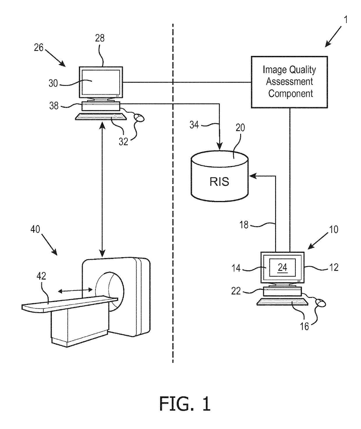
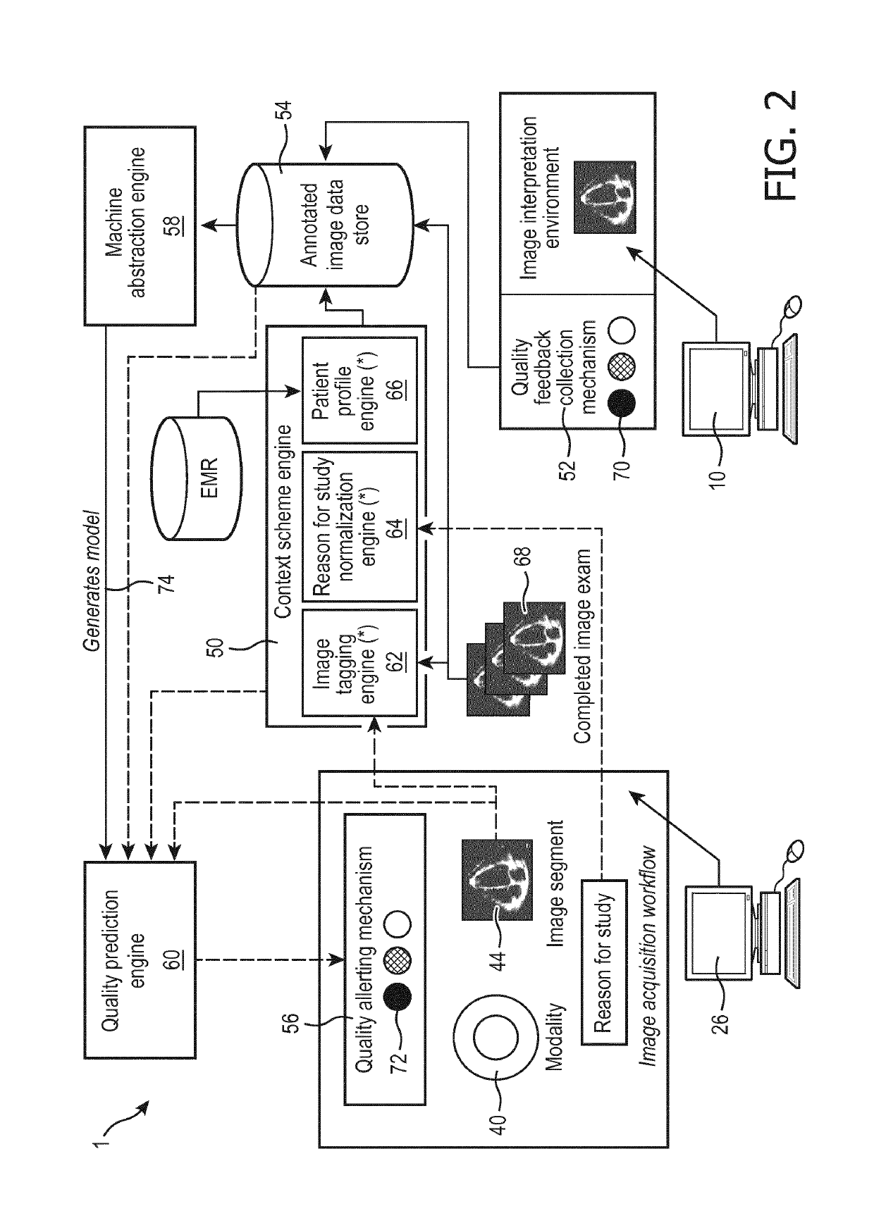
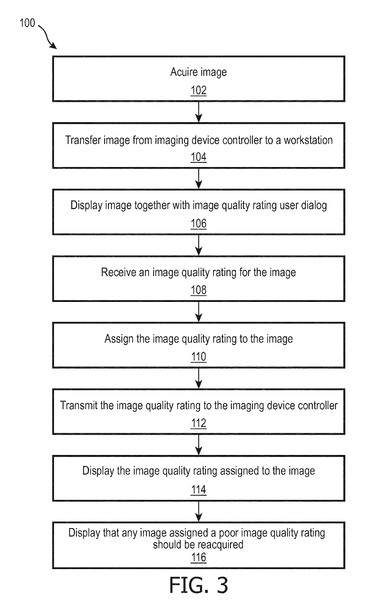
[0021]The following is generally directed to a closed-loop system that provides an automated mechanism for assessing images at the time of acquisition. In this way, the technologist or sonographer is alerted if the images are not of sufficient quality and can acquire new images while the patient is still at the imaging facility.

[0022]To this end, a medical workstation is modified to provide a tool by which the image interpreter grades quality of the images being read. As the image interpreter typically carries a heavy workload, this tool should preferably make it simple for the image interpreter to provide feedback in one embodiment the image interpreter is asked to make a selection: “Good”, “Fair”, or “Poor” and the images are so labeled. In this way a training dataset is efficiently collected, comprising actual medical images graded as to image quality by actual image interpreters qualified to perform such grading.

[0023]The training data are used to train a classifier (i.e. machin...


PUM


Abstract

A medical imaging apparatus includes a radiology workstation (10) with a workstation display (14) and one or more workstation user input devices (16). A medical imaging device controller (26) includes a controller display (30) and one or more controller user input devices (32). The medical imaging device controller is connected to control a medical imaging device (40) to acquire medical images (44). One or more electronic processors (22, 38) are programmed to: operate the medical workstation to provide a graphical user interface (GUI) (24) that displays medical images stored in a radiology information system (RIS) (20), receives entry of medical examination reports, displays an image rating user dialog (70), and receives, via the image rating user dialog, image quality ratings for medical images displayed at the medical workstation; operate the medical imaging device controller to perform an imaging examination session including operating the medical imaging device controller to control the medical imaging device to acquire session medical images; while performing the imaging examination session, assign quality ratings to the session medical images based on image quality ratings received via the image quality rating user dialog displayed at the medical workstation; and while performing the imaging examination session, display quality ratings assigned to the session medical images.

Description

FIELD[0001]The following relates to the medical arts, medical acquisition arts, medical reporting arts and related arts.BACKGROUND[0002]Medical imaging is typically performed in two phases; image acquisition and image interpretation. The acquisition is performed by technologists (or sonographers for ultrasound), who are technically trained but are not generally qualified to perform medical diagnosis based on the images. The image interpreter, oncologist, or other medical professional performs the medical diagnosis, usually at a later time (e.g. the next day or even a few days after the imaging acquisition). As a consequence, the technologist or sonographer sometimes acquire images that turn out to be diagnostically non-optimal or even non-diagnostic (i.e. the image interpreter is unable to draw diagnostic conclusions based on image acquisition deficiencies).[0003]There is an increasing emphasis on reducing costs in medicine, including medical imaging. As a consequence, appropriatene...

Claims


Application Information

Patent Timeline
IPC IPC(8): A61B6/00G06T7/00G06N3/08G06N20/00G06V10/764G06V10/82
CPCA61B6/5205A61B6/54G06T7/0012G06T2207/20081G06N20/00G06T2207/10081G06T2207/20084G06N3/08G16H30/40G16H30/20G16H40/63G06V10/993G06V10/82G06V10/764G06F18/24143
Inventor SEVENSTER, MERLIJNQIAN, YUECHENJIA, YUGANGTAHMASEBI MARAGHOOSH, AMIR MOHAMMAD
Owner KONINKLJIJKE PHILIPS NV
Why Eureka
Try Eureka