
Method and apparatus for image processing and visualization for analyzing cell kinematics in cell culture

a cell culture and cell kinematic technology, applied in image enhancement, instruments, monitoring particle agglomeration, etc., can solve the problems of low signal-to-noise ratio, low signal-to-noise ratio, uneven background illumination, etc., and achieve segmented images of low contras

Active Publication Date: 2019-05-02
CHUNG YUAN CHRISTIAN UNIVERSITY
View PDF2 Cites 11 Cited by
  • Summary
  • Abstract
  • Description
  • Claims
  • Application Information

AI Technical Summary

Benefits of technology

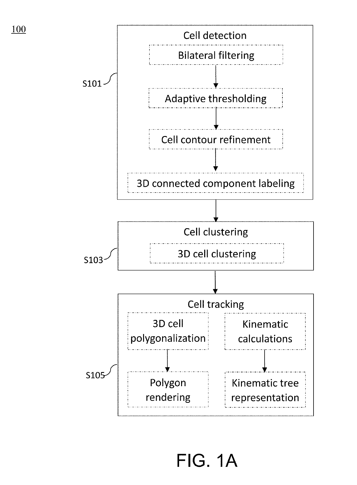
The present patent describes a method for analyzing cell kinematics in a nucleated cell culture. The method automatically detects and tracks nucleated cells and colonies from a time-series sequence of fluorescence microscopic images. The method uses a simplified approach to identify and track cells based on their nucleus and cell cluster. The method can quantitatively analyze the kinematics of cells and colonies from the fluorescence microscopic images. The method can be used to study cell behavior and cell fate in a variety of cell cultures.

Problems solved by technology

However, to analyze cell kinematics (such as, cell motion, reproduction, diffusion, and attachment), a massive number of time-series images would be taken, and therefore, the interpretation of these data is quite tedious and time-consuming.
Be that as it may, tracking fluorescent cells faces challenges of non-homogenous staining, low signal-to-noise ratio, uneven background illumination, and photobleaching.
Nevertheless, the segmented images are usually of low contrast and poor depth resolutions due to the tight packing of cells.
Although these methods may yield the segmentation of cell nuclei in microscopic images, the detection results are often limited in terms of 3D visualization of cells and / or colonies.

Method used


Image

  • Method and apparatus for image processing and visualization for analyzing cell kinematics in cell culture
  • Method and apparatus for image processing and visualization for analyzing cell kinematics in cell culture
  • Method and apparatus for image processing and visualization for analyzing cell kinematics in cell culture

Examples


example 2

[0165](1) Embryonic Stem (ES) Cell Formation

[0166]The mouse embryonic stem cells were cultured following the protocols set forth above in Example 1.

[0167](2) Imaging Conditions

[0168]The present examples used time-lapse fluorescence microscopic images. A cross-sectional image was acquired by focusing on the microscope lens. The cross-sectional images of each time frame (t=0, 1, 2, . . . ) were obtained by moving the focusing position as using a confocal microscopy. The 3D time-series images at certain time frame could be considered as constituting a volume data. Meanwhile, 2D time-series images could be time-lapse cross-sectional images at a constant cross-section or perspective images as using a wide-field microscope.

[0169]In this example, a set of 3D time-series images obtained from live Hela cells and a set of 2D time-series perspective images obtained from live mouse ES cells were studied. These data were captured at RIKEN BioResource center, Tsukuba, Japan. FIG. 9 shows two 2D f...

example 3

[0182](1) Embryonic Stem (ES) Cell Formation

[0183]The mouse embryonic stem cells were cultured following the protocols set forth above in Example 1.

[0184](2) Imaging Conditions

[0185]In this study, the CV-1000 (Yokogawa) confocal microscope was used to obtain fluorescence microscopic images of embryonic stem cells. The camera being used is the model Olympus UPLSApo60xO. Table 3 summarizes the microscope setting. Three channels of fluorescent microscopic images of embryonic stem cells were obtained simultaneously. Time-lapse confocal microscopy images were taken at a time interval of 20 minutes for a total of 24-hour duration, resulting in 144 image datasets, each set contained 9 slices of images with a thickness of 3.75 μm, and each slice image was with the resolution of 512×512 pixels and each pixel was approximately 0.26 μm.

TABLE 3Channel NameChannel 1Channel 2Channel 3Excitation488 nm561 nmLampPower (%)10 30 19 EmissionBP525 / 50BP617 / 73ThroughFluorescentEGFPmRFPBright fieldExposure...


PUM

PropertyMeasurementUnit
fluorescence microscopic imagesaaaaaaaaaa
fluorescence microscopicaaaaaaaaaa
fluorescence microscopic imageaaaaaaaaaa

Abstract

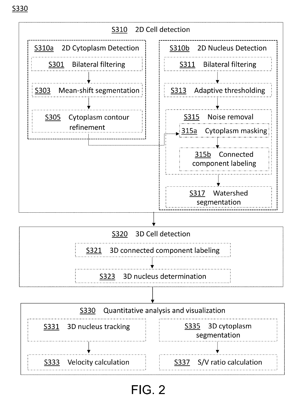
Disclosed herein are methods for analyzing cell kinematics in a nucleated cell culture from a time-series sequence of multiple fluorescence microscopic images of the nucleated cell culture. The method includes the steps of, (a) identifying every cell nucleus in each fluorescence microscopic image; (b) identifying every cell cluster using the cell nuclei identified in the step (a); and (c) tracking the cells and / or cell clusters using the cell nuclei and cell clusters identified for the fluorescence microscopic images in steps (a) and (b) respectively.

Description

STATEMENT REGARDING PRIOR DISCLOSURES BY THE INVENTOR OR A JOINT INVENTOR UNDER 37 C.F.R. 1.77(B)(6)[0001]Part of the subject matter of the invention described in the present application was published by the inventors, Yuan-Hsiang Chang, Hideo Yokota, Kuniya Abe, and Ming-Dar Tsai in an article titled “Detection and Localization of Mouse Induced Pluripotent Stem Cell Formation using Time-Lapse Fluorescence Microscopy Images,” which was disclosed during the 2016 IEEE 16th International Conference on Biolnformatics and BioEngineering (BIBE) held between Oct. 31 to Nov. 2, 2016. Part of the subject matter of the invention described in the present application was published by the inventors, Yuan-Hsiang Chang, Hideo Yokota, Kuniya Abe, Chia-Tong Tang, and Ming-Dar in an article titled “Automated Detection and Tracking of Cell Clusters in Time-Lapse Fluorescence Microscopy Images,” which was first published online by Journal of Medical and Biological Engineering on Jan. 17, 2017. Part of ...

Claims


Application Information

Patent Timeline
Patent Type & Authority Applications(United States)
IPC IPC(8): G06K9/00G01N15/14G01N15/02G06T7/00G06T7/207G06T7/60G06T7/35G06T7/55G06T7/11
CPCG06K9/00127G01N15/1468G01N15/02G06T7/0002G06T7/207G06T7/60G06T7/35G06T7/55G06T7/11G01N2015/0003G01N2015/0065G01N2015/0092G01N2015/025G01N2015/1075G01N2015/1087G01N2015/1093G06T2207/10056G06T2207/30024G01N2015/1006G06T7/136G06T7/187G06T7/155G06T2207/20028G06T7/277G06V20/69G01N2015/1027G01N2015/1029G01N2015/103G01N15/1433G01N15/01
Inventor CHANG, YUAN-HSIANGYOKOTA, HIDEOABE, KUNIYATSAI, MING-DAR
Owner CHUNG YUAN CHRISTIAN UNIVERSITY
Learn More