
System and methods for at-home ultrasound imaging

a technology of ultrasound imaging and at-home use, applied in the field of medical imaging, can solve problems such as burdening patients, and achieve the effect of reducing the number of in-clinic medical imaging sessions

Inactive Publication Date: 2018-12-06
GENERAL ELECTRIC CO
View PDF5 Cites 22 Cited by
  • Summary
  • Abstract
  • Description
  • Claims
  • Application Information

AI Technical Summary

Benefits of technology

The patent describes a system that uses a position sensor to help a probe identify its current position relative to a target position. This allows the probe to acquire image data in the target scan plane. The system also provides feedback to the user to move the probe to the target position, which can help non-clinical users perform ultrasound imaging at home and reduce the number of in-clinic medical imaging sessions.

Problems solved by technology

These frequent medical imaging sessions may place a burden on the patient.

Method used


Image

  • System and methods for at-home ultrasound imaging
  • System and methods for at-home ultrasound imaging
  • System and methods for at-home ultrasound imaging

Examples


Embodiment Construction

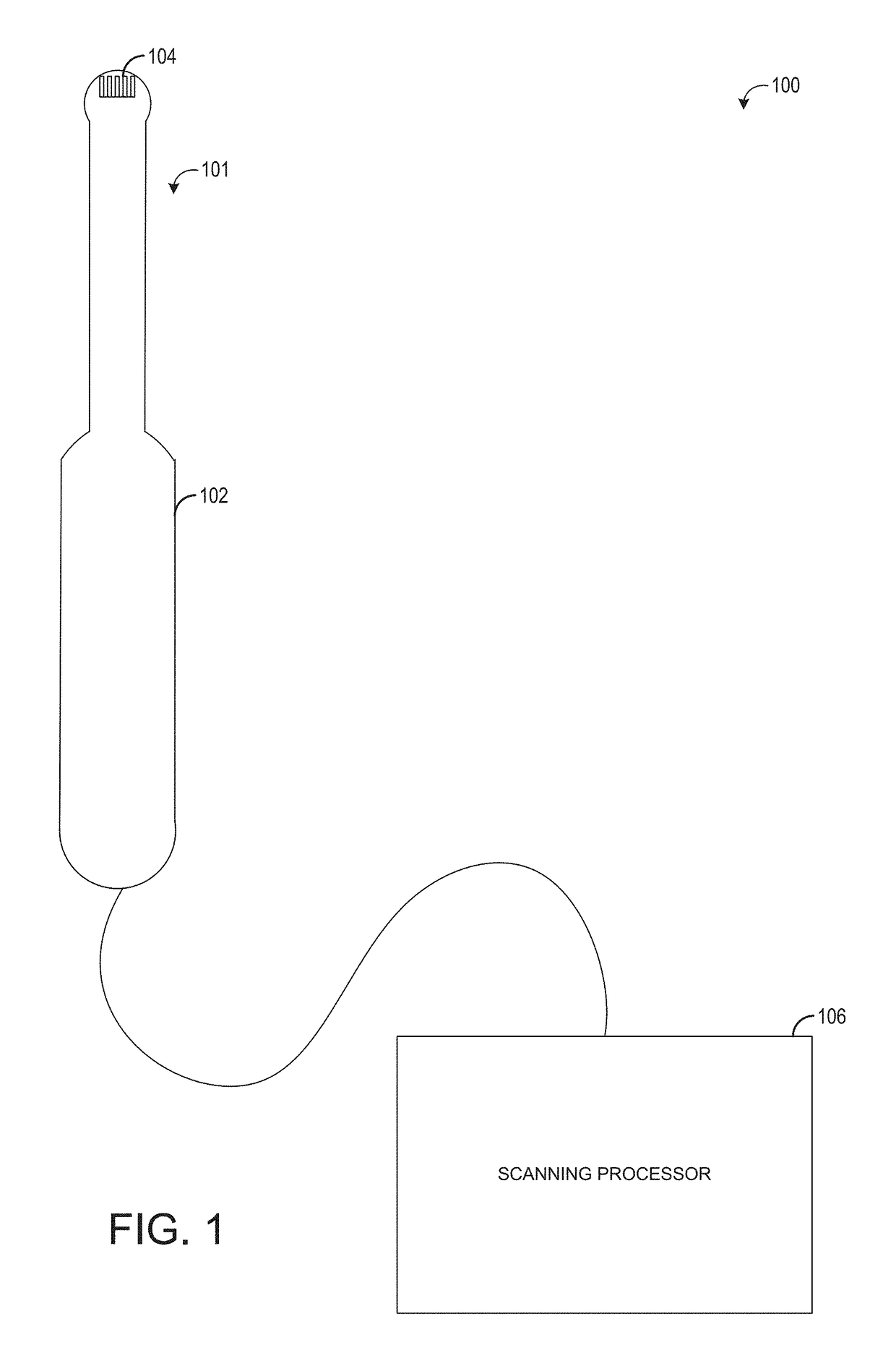
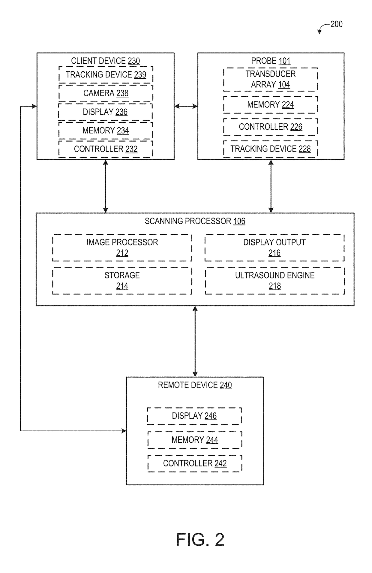
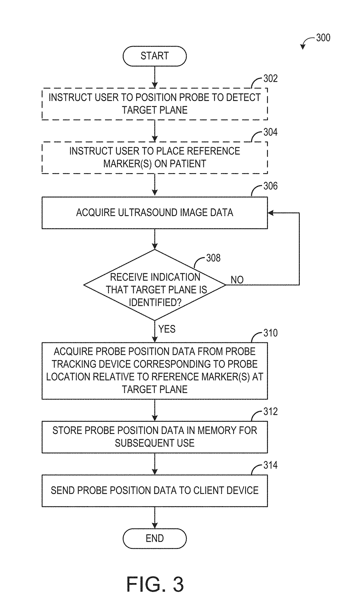
[0013]The following description relates to various embodiments of an at-home ultrasound system, which may be used during in vitro fertilization (IVF) monitoring. The at-home ultrasound system may include a transvaginal (TV) ultrasound probe, such as the probe illustrated in FIG. 1. In one example, the TV probe may include an ultrasound transducer and a position tracking system that may include one or more position sensors to track the location of the probe, such as the sensors depicted in FIG. 2. The probe may be used to provide semi-automated at-home ultrasound imaging. For example, the sensor information may be used to instruct an operator to position the probe at one or more target locations during the at-home ultrasound imaging. The image information acquired during the imaging may be used to construct a three-dimensional volume from which images may be generated and sent to a remote computing device (e.g., accessible by a clinician) for evaluation. Example methods for performin...


PUM


Abstract

Various methods and systems are provided for a guided at-home ultrasound imaging session. In one example, a system for ultrasonically scanning a tissue sample includes a hand-held ultrasound probe including a transducer array of transducer elements, a probe position tracking device including one or more position sensors coupled to the ultrasound probe, and a controller. The controller is configured to, during an imaging session, determine a position of the ultrasound probe relative to a target position based on position data collected by the probe position tracking device, and responsive to an indication that the ultrasound probe is at the target position, acquire image data with the transducer array.

Description

FIELD[0001]Embodiments of the subject matter disclosed herein relate to medical imaging and the facilitation of ultrasonic tissue scanning.BACKGROUND[0002]Certain medical procedures, such as in vitro fertilization, may include multiple, frequent medical imaging sessions in order to monitor the progress of the procedure. These frequent medical imaging sessions may place a burden on the patient.BRIEF DESCRIPTION[0003]In one embodiment, a system for ultrasonically scanning a tissue sample includes a hand-held ultrasound probe comprising a transducer array of transducer elements, a probe position tracking device including one or more position sensors coupled to the ultrasound probe, and a controller. The controller is configured to, during an imaging session, determine a position of the ultrasound probe relative to a target position based on position sensor data collected by the one or more position sensors during an imaging session, and responsive to an indication that the ultrasound p...

Claims


Application Information

Patent Timeline
Patent Type & Authority Applications(United States)
IPC IPC(8): A61B8/00A61B5/00G06F19/00
CPCA61B8/4427A61B8/4254A61B5/4848G06F19/3418A61B8/0858G06F19/321A61B8/5223A61B8/4444A61B8/565A61B8/582G16H40/63
Inventor MIENKINA, MARTIN PAULSCHWAB, FIONA
Owner GENERAL ELECTRIC CO
Learn More