Calculation of perfusion parameters in medical imaging

a perfusion parameter and imaging technology, applied in the field of finding perfusion parameters from medical images, can solve the problems of extremely heterogeneous hemodynamics and probably not a good assumption

Active Publication Date: 2018-03-22
PHILIPS MEDICAL SYST TECH
View PDF0 Cites 7 Cited by
  • Summary
  • Abstract
  • Description
  • Claims
  • Application Information

AI Technical Summary

Benefits of technology

[0034]For example, hardware for performing selected tasks according to embodiments of the invention could be implemented as a chip or a circuit. As software, selected tasks according to embodiments of the invention could be implemented as a plurality of software instructions being executed by a computer using any suitable operating system. In an exemplary embodiment of the invention, one or more tasks according to exemplary embodiments of method and / or system as described herein are performed by a data processor, such as a computing platform for executing a plurality of instructions. Optionally, the data processor includes a volatile memory for storing instructions and / or data and / or a non-volatile storage, for example, a magnetic hard-disk and / or removable media, for storing instructions and / or data. Optionally, a network connection is provided as well. A display and / or a user input device such as a keyboard or mouse are optionally provided as well.

Problems solved by technology

Given that tumor hemodynamics are extremely heterogeneous across and within the same tumor, this is probably not a good assumption.

Method used


Image

  • Calculation of perfusion parameters in medical imaging
  • Calculation of perfusion parameters in medical imaging
  • Calculation of perfusion parameters in medical imaging

Examples


Embodiment Construction

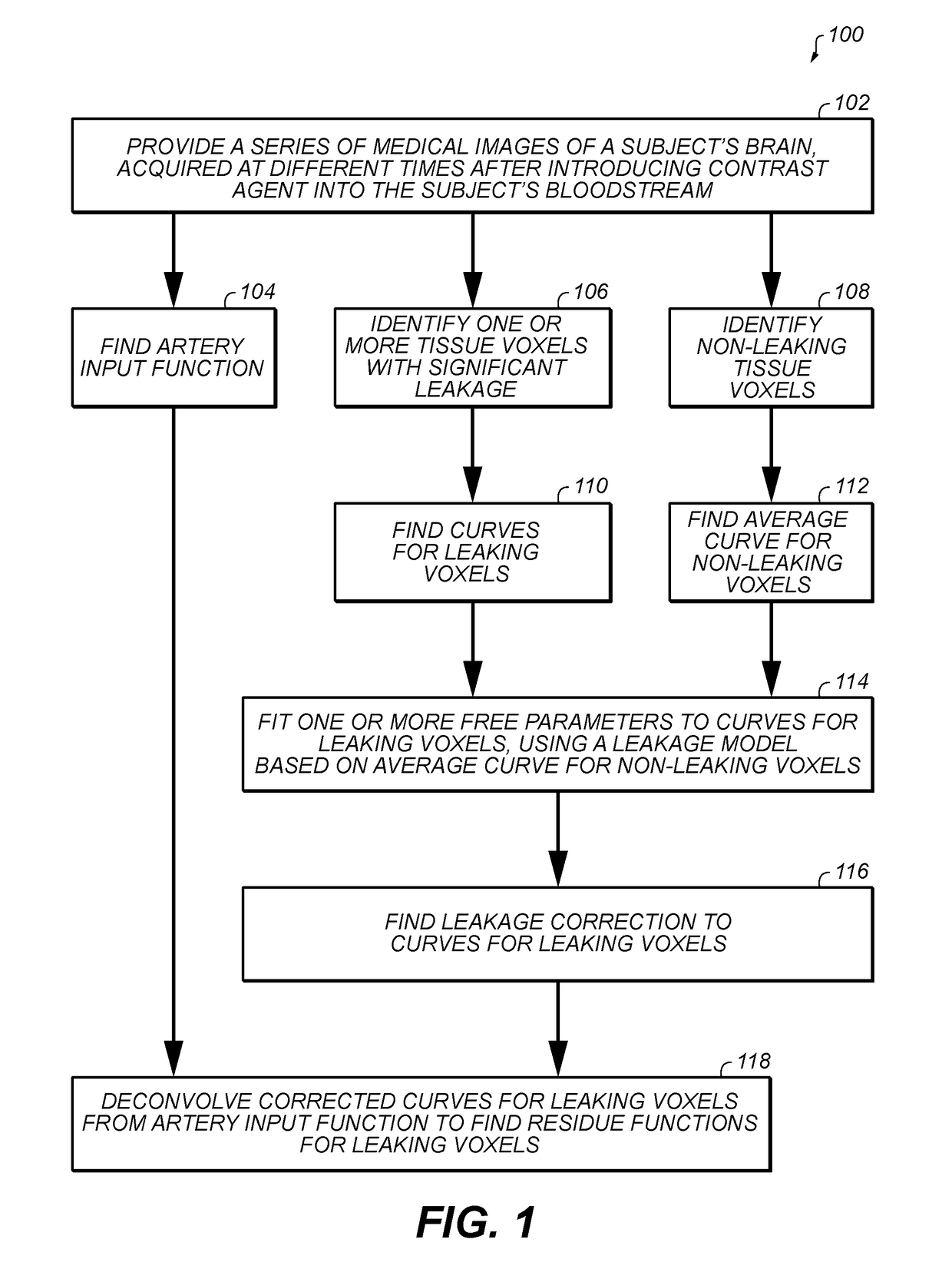
[0046]The present invention, in some embodiments thereof, relates to a method of finding perfusion parameters from medical images and, more particularly, but not exclusively, to a method of finding cerebral perfusion parameters from MRI or CT images of the brain.

[0047]An aspect of some embodiments of the invention concerns a method of finding a perfusion residue function, at different positions in brain tissue of a subject, using data derived from a series of medical images of the tissue acquired at a plurality of successive times after introducing a contrast agent into the subject's bloodstream, and correcting the measured concentration of contrast agent in the capillaries at each voxel for leakage of the contrast agent into the extravascular extracellular space (EES). The method first obtains a signal as a function of time for concentration of contrast agent in the capillaries averaged over voxels that do not exhibit leakage, and initially assumes that the same function applies to...


PUM


Abstract

A method of determining a residue function in brain tissue, from medical images acquired after introducing contrast agent into the blood, correcting for contrast agent leakage into the tissue, comprising: a) providing time signals indicating contrast agent concentration for leaking voxels, a time signal indicating average contrast agent concentration for non-leaking voxels, and an artery input function, all derived from the images; b) fitting the leaking voxel signals to a model time signal with a free parameter for leakage rate, the model assuming that the concentration of contrast agent perfusing through a leaking voxel has a same shape as a function of time as the average contrast agent concentration for non-leaking voxels; c) using the best fit leakage rate parameter to make a correction for leakage to the leaking voxel signals; and d) deconvolving the corrected signals from the artery input function, to find the residue function.

Description

CROSS REFERENCE TO RELATED APPLICATIONS[0001]This application claims the benefit of U.S. Provisional 62 / 397,993, provisionally filed on Sep. 22, 2016, entitled “CALCULATION OF PERFUSION PARAMETERS IN DSC MRI IMAGING,” in the name of Ohad SILBERT, incorporated herein by reference as if fully set forth in their entirety.FIELD AND BACKGROUND OF THE INVENTION[0002]The present invention, in some embodiments thereof, relates to a method of finding perfusion parameters from medical images and, more particularly, but not exclusively, to a method of finding cerebral perfusion parameters from MRI or CT images of the brain.[0003]Various medical imaging modalities can be useful for measuring rates of perfusion of body tissue as a function of position in the tissue, to detect the precise location and extent of deficiencies in perfusion. Such methods are important for evaluating brain tumors, which are often associated with deficiencies in perfusion, and with leakage of blood or components of blo...

Claims


Application Information

Patent Timeline
Patent Type & Authority Applications(United States)
IPC IPC(8): A61B6/00A61B6/03A61B5/026A61B5/00A61B5/0295G01R33/56G01R33/563G06T7/11G06T7/00
CPCA61B6/5258A61B6/032A61B5/0263A61B6/507A61B6/5217A61B5/0042A61B6/501A61B5/0295A61B5/7203A61B5/7278G01R33/5601G01R33/56366G06T7/11G06T7/0016G06T2207/30016G06T2207/30104G06T2207/10081G06T2207/10096A61B5/055A61B6/481A61B2576/026G16H50/30
Inventor SILBERT, OHAD
Owner PHILIPS MEDICAL SYST TECH
Why Eureka
Try Eureka