
Image processor, image processing method, and endoscope system

a technology of image processing and endoscopy, which is applied in the field of image processing, image processing method, and endoscopy system, can solve the problems of complex diagnosis in consideration of a plurality of items of blood vessel information

Active Publication Date: 2017-09-14
FUJIFILM CORP
View PDF6 Cites 6 Cited by
  • Summary
  • Abstract
  • Description
  • Claims
  • Application Information

AI Technical Summary

Benefits of technology

The invention provides an image processor, method, and endoscope system that use blood vessel information to calculate blood vessel parameters and support doctors' diagnosis. The system can extract blood vessels from an endoscopic image and calculate various parameters such as the number of branches, branch angle, distance between branch points, thickness, change in thickness, degree of complexity of change in thickness, length, intervals, depth with a mucous membrane as a reference, height difference, inclination, area, density, contrast, color, change in color, the degree of meandering, blood concentration, the degree of oxygen saturation, the percentage of arteries, the percentage of veins, the concentration of an administered pigment, a traveling pattern, and blood flow rate. The system can also determine the state of the mucous membrane of the observation object using the blood vessel parameters. Overall, the invention provides a more accurate and reliable tool for diagnosis and treatment.

Problems solved by technology

However, doctors do not perform diagnosis on the basis of one kind of blood vessel information out of the blood vessel information, such as the depth, thickness, and density of the blood vessels, and the diagnosis indexes obtained from the color balance of pixels representing the blood vessels, but complexly perform diagnosis in consideration of a plurality of items of blood vessel information, including blood vessel information that is not evaluated by related-art endoscope systems other than the above blood vessel information.

Method used


Image

  • Image processor, image processing method, and endoscope system
  • Image processor, image processing method, and endoscope system
  • Image processor, image processing method, and endoscope system

Examples


Embodiment Construction

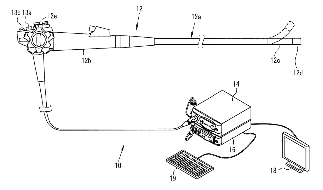
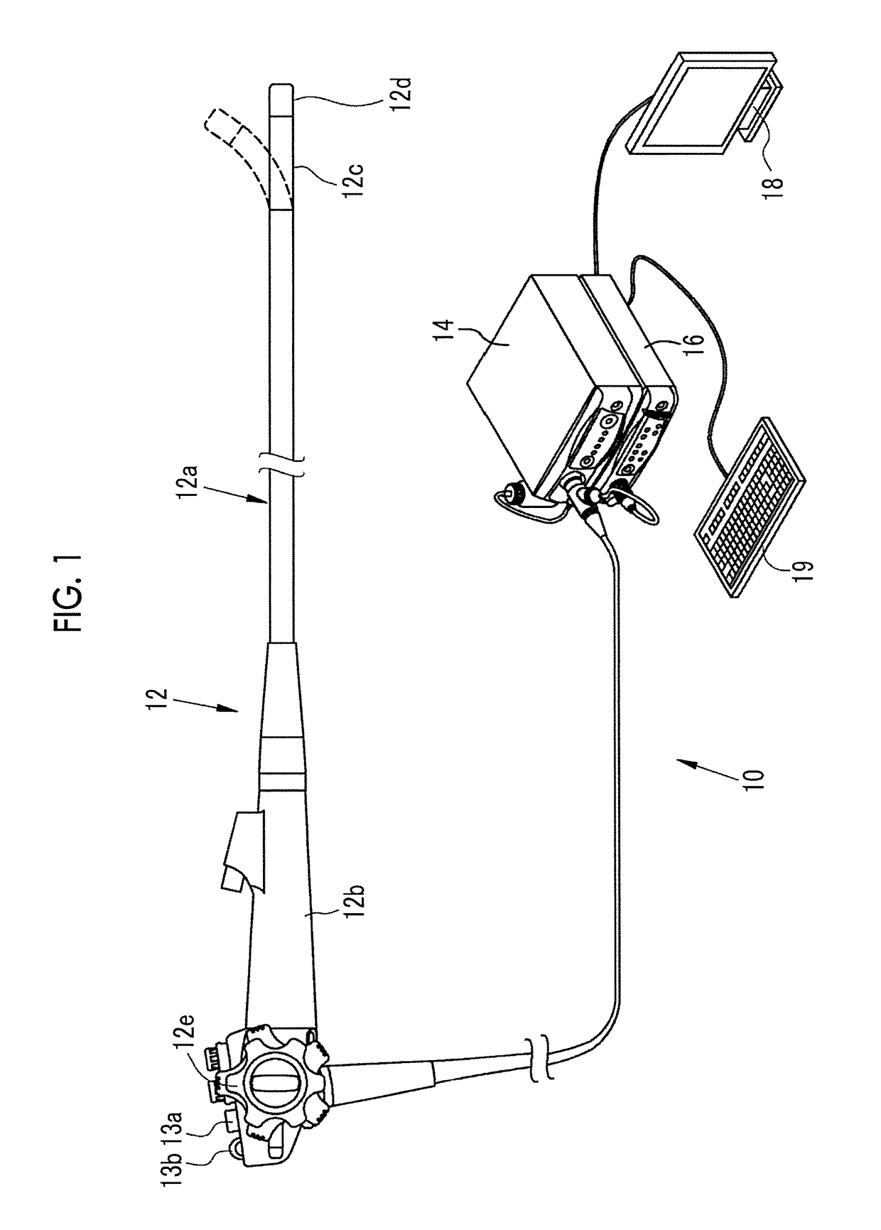
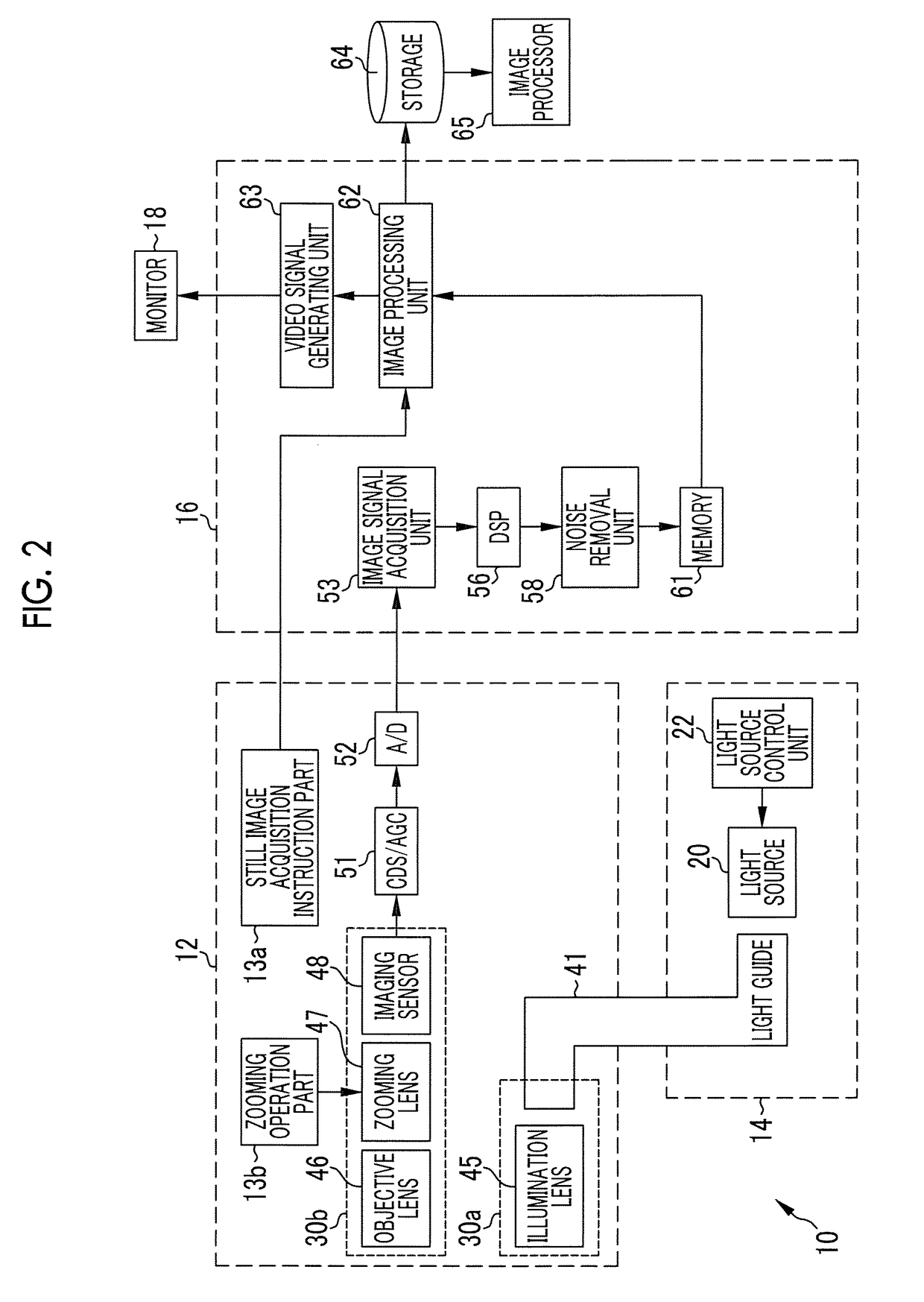
[0035]As illustrated in FIG. 1, an endoscope system 10 has an endoscope 12, a light source device 14, a processor device 16, a monitor 18, and a console 19. The endoscope 12 is optically connected to the light source device 14, and is electrically connected to the processor device 16. The endoscope 12 has an insertion part 12a to be inserted into a subject, an operating part 12b provided at a base end portion of the insertion part 12a, and a bending part 12c and a distal end part 12d provided on a distal end side of the insertion part 12a. By operating an angle knob 12e of the operating part 12b, the bending part 12c makes a bending motion. The distal end part 12d is directed in a desired direction by this bending motion.

[0036]Additionally, the operating part 12b is provided with a still image acquisition instruction part 13a and a zooming operation part 13b other than the angle knob 12e. The still image acquisition instruction part 13a is used in order to input a still image acquis...


PUM


Abstract

An image processor calculates diagnosis support parameters on the basis of the state of blood vessels of an observation object. The image processor includes an image acquisition unit that acquires an endoscopic image, a blood vessel extraction unit that extracts blood vessels, using the endoscopic image, a blood vessel information calculation unit, and a blood vessel parameter calculation unit. The blood vessel information calculation unit calculates a plurality of items of blood vessel information including at least two or more among the number of pieces, the number of branches, a branch angle, a distance between branch points, the number of intersections, thickness, a change in thickness, the degree of complexity of a change in thickness, length, intervals, depth with a mucous membrane as a reference, and the like. The blood vessel parameter calculation unit calculates blood vessel parameters by calculating using the plurality of items of blood vessel information.

Description

CROSS-REFERENCE TO RELATED APPLICATION[0001]This application is a Continuation of PCT International Application PCT / JP2016 / 052323 filed on 27 Jan. 2016, which claims priority under 35 USC 119(a) from Japanese Patent Application No. 2015-015179 filed on 29 Jan. 2015, and Japanese Patent Application No. 2015-166101 filed on 25 Aug. 2015. The above application is hereby expressly incorporated by reference, in its entirety, into the present application.BACKGROUND OF THE INVENTION[0002]1. Field of the Invention[0003]The present invention relates to an image processor, an image processing method, and an endoscope system that calculates data, such as numerical values provided for diagnosis using an endoscopic image, using the endoscopic image captured by an endoscope.[0004]2. Description of the Related Art[0005]In the medical field, diagnosis using endoscope systems including a light source device, an endoscope, and a processor device has been performed widely. In the diagnosis using the e...

Claims


Application Information

Patent Timeline
Patent Type & Authority Applications(United States)
IPC IPC(8): A61B1/00A61B1/313A61B5/02A61B5/103A61B5/1455A61B5/1459A61B5/00A61B1/04A61B5/026
CPCA61B1/00009A61B1/04A61B1/3137A61B5/02007A61B5/0261A61B5/7278A61B5/1459A61B5/489A61B5/1032A61B5/7282A61B5/14551G02B23/24H04N7/18A61B5/0073A61B5/0084A61B2576/02A61B1/0005A61B1/041G06T7/0012G06T2207/10024G06T2207/10068G06T2207/30101G16H30/40A61B1/000094A61B1/044
Inventor KAKU, TOSHIHIKO
Owner FUJIFILM CORP
Learn More