Antigen detection using photocleavable labels
a technology of photocleavage and label, applied in the field of immunological detection using photocleavage labels, can solve the problems of limited technology, limited ability to detect individual labels, and higher order multiplexing
- Summary
- Abstract
- Description
- Claims
- Application Information
AI Technical Summary
Benefits of technology
Problems solved by technology
Method used
Image
Examples
example 1
Detection of an Antigen in a Tissue Section Using Single-Stranded Biotin-Nucleotide-Fluorophore Tags
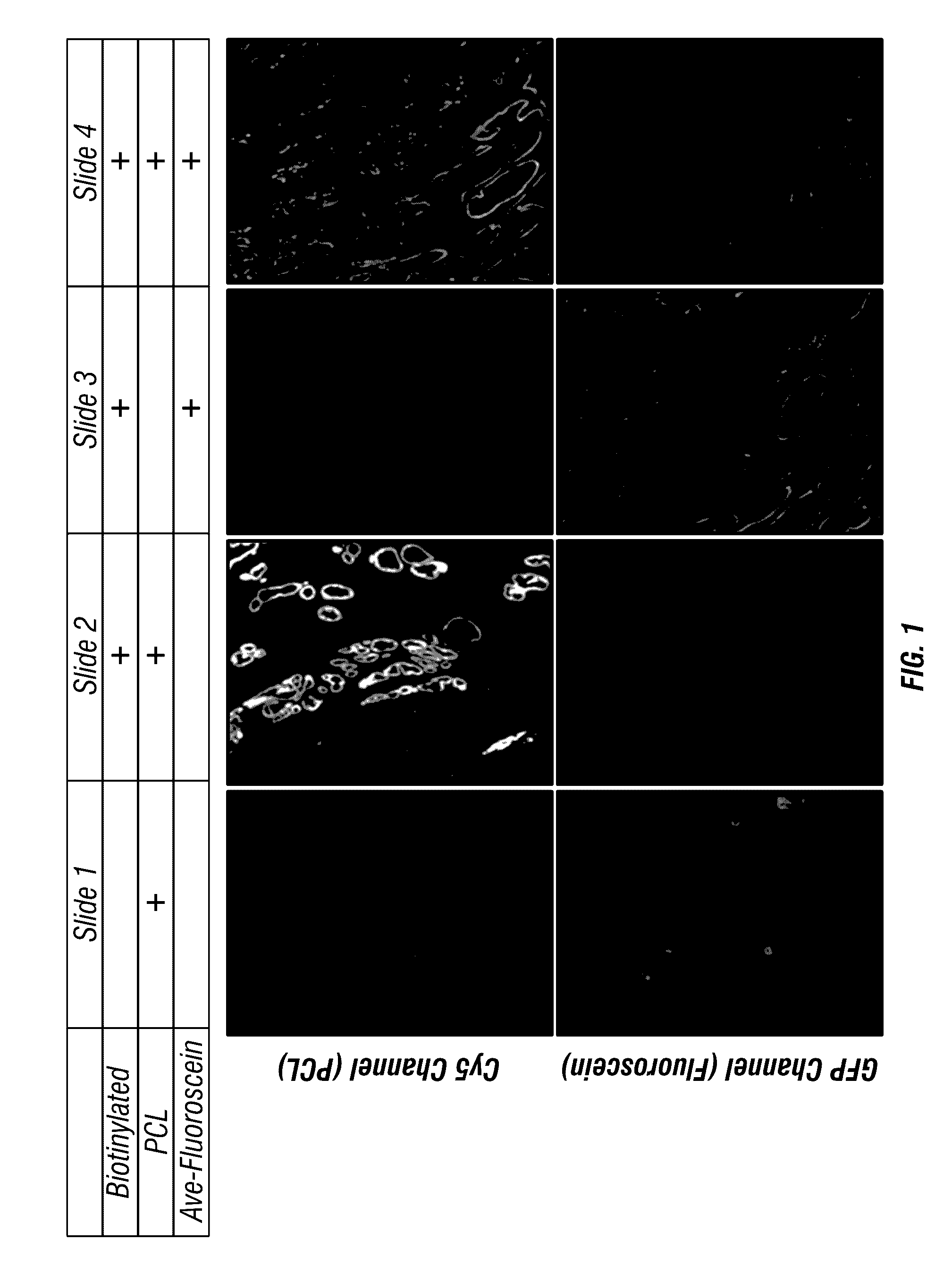
[0209]This Example illustrates an antigen-bound antigen-binding complex of formula (IX). First, formalin-fixed paraffin embedded (FFPE) prostate cancer tissue sections were prepared and blocked with BSA in PBS. Then, the sections were incubated with a mouse anti-cytokeratin antibody, followed by washing. Next, the sections were incubated with biotinylated goat anti-mouse IgG secondary antibody, followed by washing. Then, the sections were incubated with either avidin or streptavidin or buffer alone, followed by washing. Then, the sections were incubated with either a biotin-conjugated photocleavable Cy5 or avidin-fluorescein, followed by washing. The biotin-conjugated photocleavable Cy5 had a biotin modification at its 5′ end and a dC-Cy5 (FIG. 8) at its 3′ end. Finally, the sections were imaged using a 20× water immersion lens. FIG. 1 shows that the biotin-conjugated photocleavable C...
example 2
Automated Serial FFPE Tissue Staining of Two Target Proteins with Two Photocleavable Labels of Different Colors
[0211]This Example illustrates an antigen-bound antigen-binding complex of formula (VII). For serial FFPE, the tissue was first incubated with biotinylated anti-smooth muscle actin for 2 h, avidin for 30 min, and biotin-conjugated probe containing photocleavable AF532 for 20 min before the green channel was imaged. After UV exposure of 2 min at 150 mW / cm2, the tissue underwent the same staining procedure with histone H3 antibody and red photocleavable Cy5. The same field of view was imaged in the red channel and overlaid on the previous green channel using ImageJ software (FIG. 11).
[0212]Direct comparison of PCL with chemical inactivation and photobleaching of Cy dyes demonstrates the release of dye from PCL is 10-100 times more rapid. A similar comparison with other photocleavable bonds demonstrated that the PCL resulted in faster and more complete dye release than other p...
example 3
Multiplex Staining of 8 Target Regions on a Single FFPE Tissue Section with Two Color Photocleavable Labels
[0216]In another experiment, a 6 μm section of human prostate (normal) was prepared and stained in three consecutive cycles. Tissue section was probed for multiple target proteins at each cycle, a single imaging event at each cycle at various wavelengths captured signal from various target proteins. Following a cleaving event through UV exposure, the tissue was probed again for the next set of target proteins. Three such multiplex cycles resulted in capture of the following 8 targets in a single tissue section: Cycle I: Collagen (CNA35), Hoechst (nuclear DNA), anti-ALCAM and anti-CD44; Cycle II: anti-cytokeratin and anti-SMA; Cycle III: anti-Fibronectin and anti-Histone H3. Each cycle captured images from four channels: Cy7 (750 nm), Cy5 (650 nm), Cy2 (488 nm) and Hoechst (365 nm). At the completion of the experiment, the three image data sets were co-registered using the Cy2 c...
PUM
 Abstract
Description
Claims
Application Information
 - R&D
- Intellectual Property
- Life Sciences
- Materials
- Tech Scout
- Unparalleled Data Quality
- Higher Quality Content
- 60% Fewer Hallucinations
Browse by: Latest US Patents, China's latest patents, Technical Efficacy Thesaurus, Application Domain, Technology Topic, Popular Technical Reports.
© 2025 PatSnap. All rights reserved.Legal|Privacy policy|Modern Slavery Act Transparency Statement|Sitemap|About US| Contact US: help@patsnap.com


