Minimally Invasive Subgaleal Extra-Cranial Electroencephalography (EEG) Monitoring Device

a monitoring device and subgaleal technology, applied in the field of minimally invasive subgaleal extracranial electroencephalography (eeg) monitoring devices, can solve the problems of inpatient video eeg studies, high cost, and inability to detect brain electrical activity, and achieve the effect of facilitating the detection of brain electrical activity

Active Publication Date: 2017-02-09
NEW YORK UNIV
View PDF5 Cites 22 Cited by
  • Summary
  • Abstract
  • Description
  • Claims
  • Application Information

AI Technical Summary

Benefits of technology

[0004]The present invention is directed to a system, comprising an implantable body configured for implantation in a subgaleal extracranial position, the implantable body including a first electrode array including a first elongated body comprising first and second electrode contacts separated from one another by a distance selected to facilitate the detection of brain electrical activity and a unit coupled to the first electrode array. The unit includes a processor analyzing the detected brain electrical activity to determine whether an epileptic event has occurred and generating epileptic event data based on this determination and a transceiver controlled by the processor to wirelessly transmit epileptic event data to and from a remote computing device.

Problems solved by technology

Unfortunately, many patients experiencing seizures lose consciousness or are amnesic while most subtle or non-convulsive seizures may be unobserved or unrecognized.
Furthermore, unrecognized seizures increase the risk of cognitive decline, injury and death.
Video EEG studies are inpatient based and thus very expensive and only used for acute situations or for pre-surgical investigations.
Current methods for ambulatory scalp EEG recordings are not practical for long-term use and can typically be used for a maximum of 3-4 days.
These methods are also limited due to scalp EEG artifacts, battery life or the inability of patients to tolerate scalp electrodes from more than a few days.
However, not only are these highly invasive procedures expensive (generally more than $50,000), they also have a high morbidity rate.
Seizure detection systems based on body motion or motor activity, accelerometers or video detectors can only detect major convulsive events and are not implantable, limiting their value as most seizures are non-convulsive in nature and therefore not detectable with changes in motion.

Method used


Image

  • Minimally Invasive Subgaleal Extra-Cranial Electroencephalography (EEG) Monitoring Device
  • Minimally Invasive Subgaleal Extra-Cranial Electroencephalography (EEG) Monitoring Device
  • Minimally Invasive Subgaleal Extra-Cranial Electroencephalography (EEG) Monitoring Device

Examples


example i

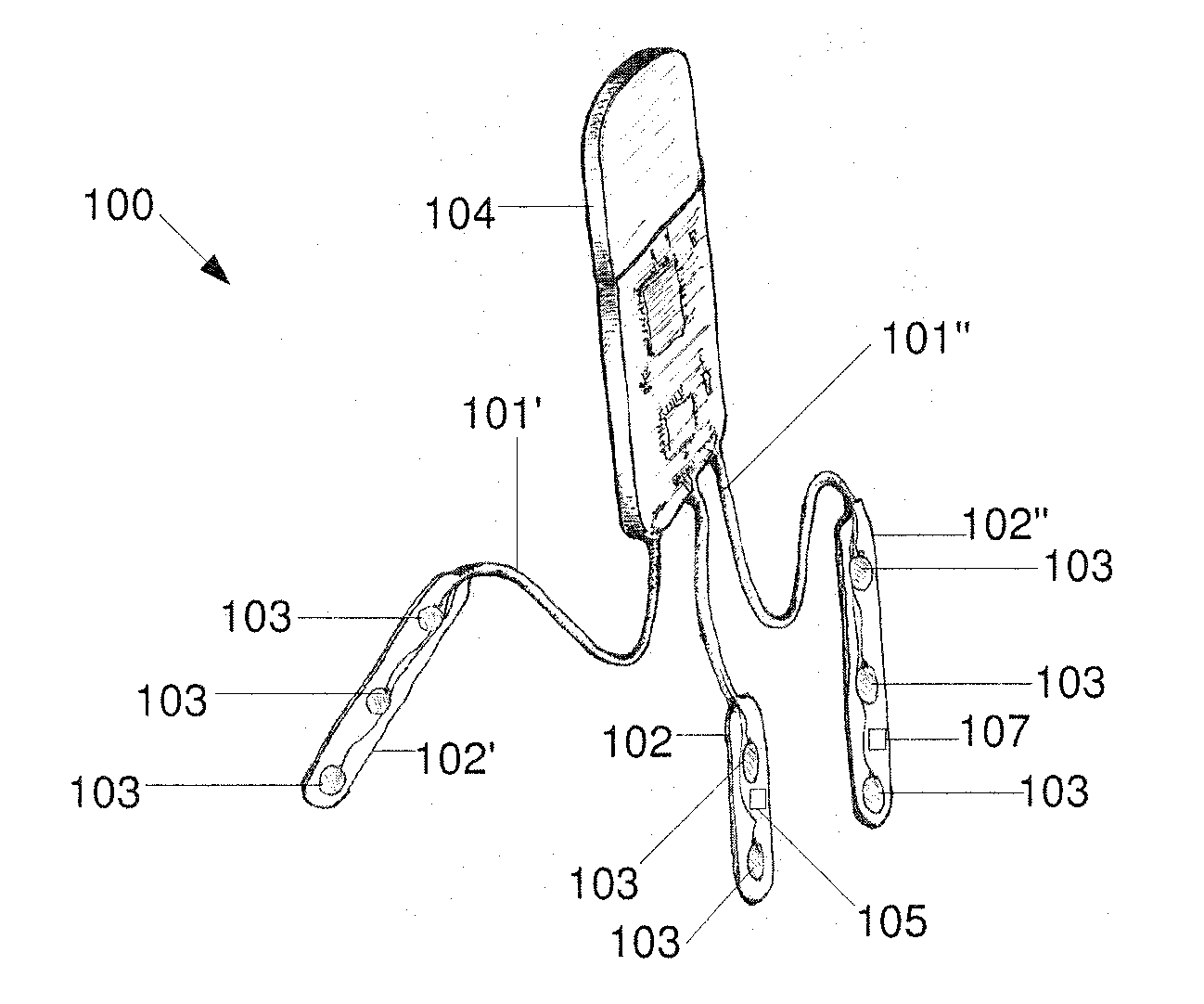
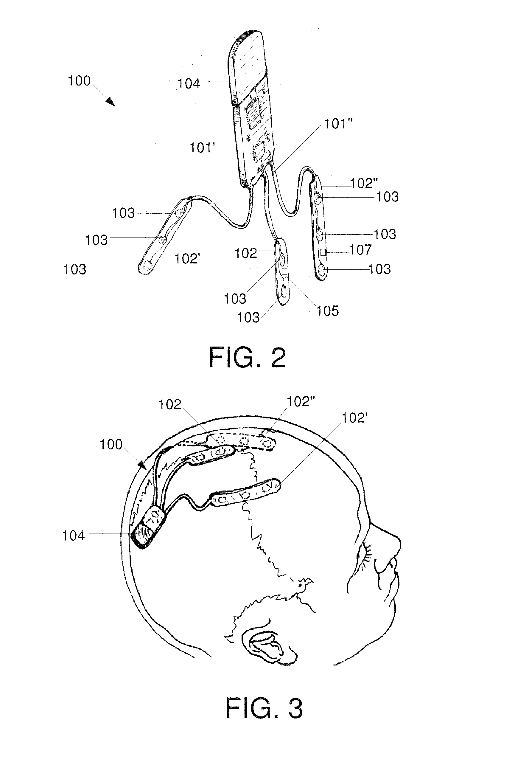
[0036]The exemplary device 100 may be implanted in a subgaleal region of a patient, near the brain, as shown in FIG. 11. As can be seen, at least one contact 103 (e.g., an electrode for recording electrophysiological data, particularly EEG data) of the device may be implanted in a subgaleal region that is between the skull 14 and the scalp 16 of the patient near the brain 12 of the patient. Specifically, the at least one contact 103 or electrode arrays 102, 102′, 102″ of the device 100 may be position perpendicular to a cranial vertex 18 of the patient. More particularly, the at least one contact 103 or electrode arrays 102, 102′, 102″ of the device 100 may lie across a mid-length of the patient's skull 14. It is contemplated that the exemplary device 200 may be alternatively implanted in the subgaleal region of the patient in the same manner described above and as shown in FIG. 11.

[0037]The device 100, 200 may be used to record EEG data from the patient extracranially without drill...


PUM


Abstract

A system includes an implantable body configured for implantation in a subgaleal extracranial position, the implantable body including a first electrode array including a first elongated body comprising first and second electrode contacts separated from one another by a distance selected to facilitate the detection of brain electrical activity and a unit coupled to the first electrode array. The unit includes a processor analyzing the detected brain electrical activity to determine whether an epileptic event has occurred and generating epileptic event data based on this determination and a transceiver controlled by the processor to wirelessly transmit epileptic event data to and from a remote computing device.

Description

[0001]This application claims priority to U.S. Provisional Patent Application Ser. No. 61 / 945,550, filed Feb. 27, 2014, the entire contents of which are hereby incorporated by reference herein.BACKGROUND[0002]For epilepsy patients, an objective seizure detection method that is safe, accurate and does not interfere with patient activities is critical to advance patient care. Seizure frequency is the most important index for determining and monitoring seizure control. Unfortunately, many patients experiencing seizures lose consciousness or are amnesic while most subtle or non-convulsive seizures may be unobserved or unrecognized. As the majority of current determinations of seizure frequency are based on patient and / or caregiver reports, these determinations represent, at best, a crude estimate of the true frequency. Studies have shown that seizure counts reported by the patients or caregivers can have an error rate as high as 60% which has important implications for therapy. Unrecogn...

Claims


Application Information

Patent Timeline
Patent Type & Authority Applications(United States)
IPC IPC(8): A61B5/0478A61B5/04A61B5/0205A61B5/053A61B5/01A61B5/11A61B5/0492A61B5/00A61B5/145A61B5/296
CPCA61B5/0478A61B5/0006A61B5/04012A61B5/6868A61B5/0533A61B5/14532A61B5/01A61B5/11A61B5/0492A61B5/02055A61B5/0002A61B5/291A61B5/296A61B5/316
Inventor KUZNIECKY, RUBEN I.DOYLE, WERNER K.PACIA, STEVEFRIEDMAN, DANIEL
Owner NEW YORK UNIV
Why Eureka
Try Eureka