Method for evaluation of function of phagocyte
a phagocyte and function technology, applied in the field of phagocyte function evaluation, can solve problems such as autologous tissue destruction, and achieve the effects of specific and convenient detection, assaying the function of phagocytes, and convenient detection
- Summary
- Abstract
- Description
- Claims
- Application Information
AI Technical Summary
Benefits of technology
Problems solved by technology
Method used
Image
Examples
example 1
Production of F1301-9-1 Antibody by Using a Synthetic Peptide for the Antigen
[0131]1-(1) Preparation of the Peptide Used for the Antigen
[0132]A peptide having the sequence defined in SEQ ID NO. 5 (corresponding to the sequence of 40th to 59th amino acid residues of rabbit CD14 amino acid sequence) having cysteine inserted at its N terminal for ligation at its end with the carrier protein via SH group was synthesized using peptide synthesizer ABI433A (Applied Biosystems). The peptide was purified by the method commonly used in the art to obtain 2 to 3 mg of the peptide.
[0133]1-(2) Preparation of Peptide Carrier Protein Using the Synthetic Peptide
[0134]The peptide prepared in 1-(1) was dissolved in distilled water to 10 mg / mL, and equal amount of this solution and 10 mg / mL Imject Maleimide Activated Keyhole Limpet Hemocyanin (KLH) (PIERCE) were mixed. The mixture was allowed to react at room temperature for 2 hours for the binding of the peptide with the carrier protein (this product ...
example 2
Preparation of F1258-7-2 Antibody by Using the Synthetic Peptide for the Antigen
[0139]2-(1) Preparation of the Peptide Used for the Antigen
[0140]A peptide having the sequence defined in SEQ ID NO. 6 (corresponding to the sequence of 1st to 30th amino acids of rabbit CD14 amino acid sequence) having cysteine inserted at its N terminal for ligation at its end with the carrier protein via SH group was synthesized using peptide synthesizer ABI433A (Applied Biosystems). The peptide was purified by the method commonly used in the art to obtain 2 to 3 mg of the peptide.
[0141]2-(2) Preparation of Peptide Carrier Protein Using the Synthetic Peptide
[0142]SEQ ID NO. 6 peptide—KLH and SEQ ID NO. 6 peptide—BSA were prepared by the method similar to that of to 1-(2).
[0143]2-(3) Production of Hybridoma Clone by Using the Synthetic Peptide for the Antigen
[0144]Clone F1258-7-2 producing the monoclonal antibody for the SEQ ID NO. 6 peptide prepared in 2-(2) was produced by the method similar to that ...
example 3
Preparation of the Assay System for Rabbit sCD14-ST
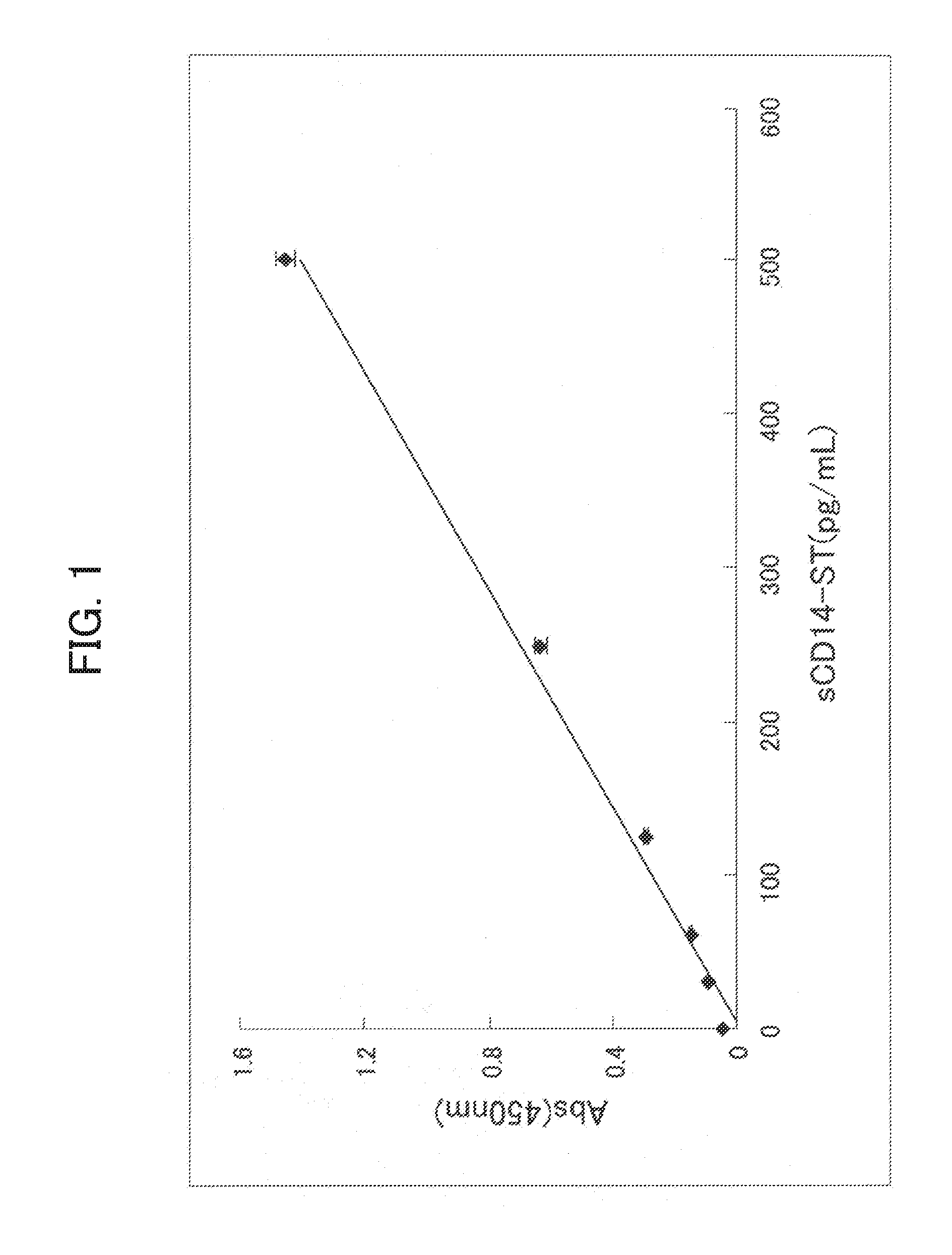
[0147]In order to prepare a system capable of specifically detecting of the rabbit sCD14-ST, a sandwich ELISA system was prepared by using the antibodies prepared in Example 1-(4) and Example 2-(4).
[0148]3-(1) Preparation of F1258-7-2 Fab′-HRP
[0149]To prepare F(ab′)2 of the F1258-7-2 antibody, the purified F1258-7-2 antibody prepared in Example 2-(4) was treated with pepsin. More specifically, the purified F1258-7-2 antibody was subjected to buffer exchange with 100 mM acetate buffer (pH 4.4) containing 2M urea, and pepsin (Boehringer) was added so that antibody: enzyme was 30:1 (weight ratio). After the addition, the reaction was promoted at 37° C. for 6 hours. At the completion of the reaction, 1M Tris-hydrochloric acid buffer (pH 8.0) was added to bring the pH back to approximately neutral. Next, F(ab′)2 was purified. More specifically, the antibody treated with pepsin was applied to Prosep G (Millipore) to remove the Fc moiety a...
PUM
 Abstract
Description
Claims
Application Information
 - R&D
- Intellectual Property
- Life Sciences
- Materials
- Tech Scout
- Unparalleled Data Quality
- Higher Quality Content
- 60% Fewer Hallucinations
Browse by: Latest US Patents, China's latest patents, Technical Efficacy Thesaurus, Application Domain, Technology Topic, Popular Technical Reports.
© 2025 PatSnap. All rights reserved.Legal|Privacy policy|Modern Slavery Act Transparency Statement|Sitemap|About US| Contact US: help@patsnap.com


