Supercharge Your Innovation With Domain-Expert AI Agents!

Image processing method to diagnose cutaneous lesion, diagnostic apparatus used for the same method, and medium storing program associated with the same method

Active Publication Date: 2016-09-22
CASIO COMPUTER CO LTD
View PDF2 Cites 11 Cited by
  • Summary
  • Abstract
  • Description
  • Claims
  • Application Information

AI Technical Summary

Benefits of technology

The patent describes a method for processing a cutaneous image to diagnose a cutaneous lesion. The method involves obtaining a first detail image and a second detail image with different properties, and generating a third detail image based on the combination of the first and second detail images. This third detail image is then used to create a new base image to restore the original brightness component and generate a highlighted image. The technical effect is improved image processing for accurate diagnosis of cutaneous lesions.

Problems solved by technology

However, not only discrimination between a mole and a spot but also discrimination between a benign tumor and a malignant tumor are substantially difficult with a naked eye inspection and even a magnifying glass inspection.

Method used


Image

  • Image processing method to diagnose cutaneous lesion, diagnostic apparatus used for the same method, and medium storing program associated with the same method
  • Image processing method to diagnose cutaneous lesion, diagnostic apparatus used for the same method, and medium storing program associated with the same method
  • Image processing method to diagnose cutaneous lesion, diagnostic apparatus used for the same method, and medium storing program associated with the same method

Examples


application example 1

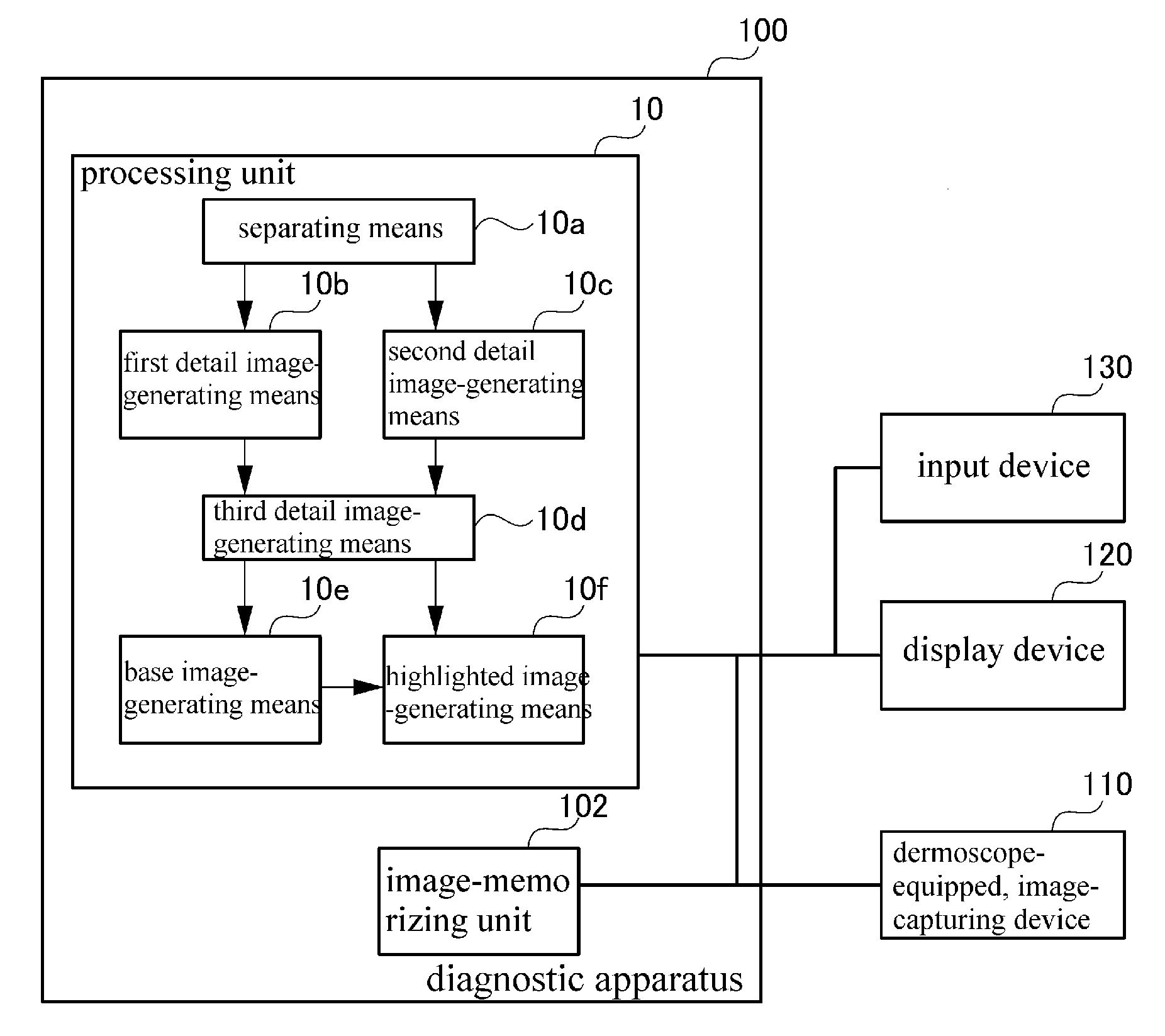
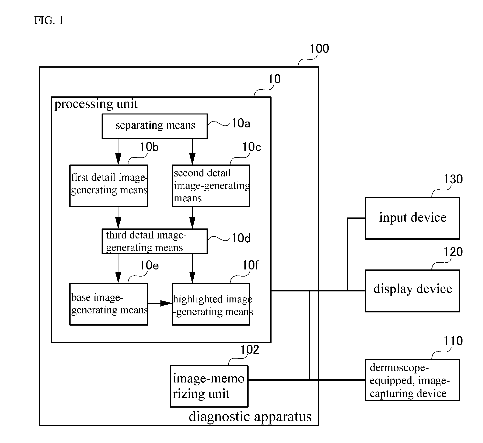
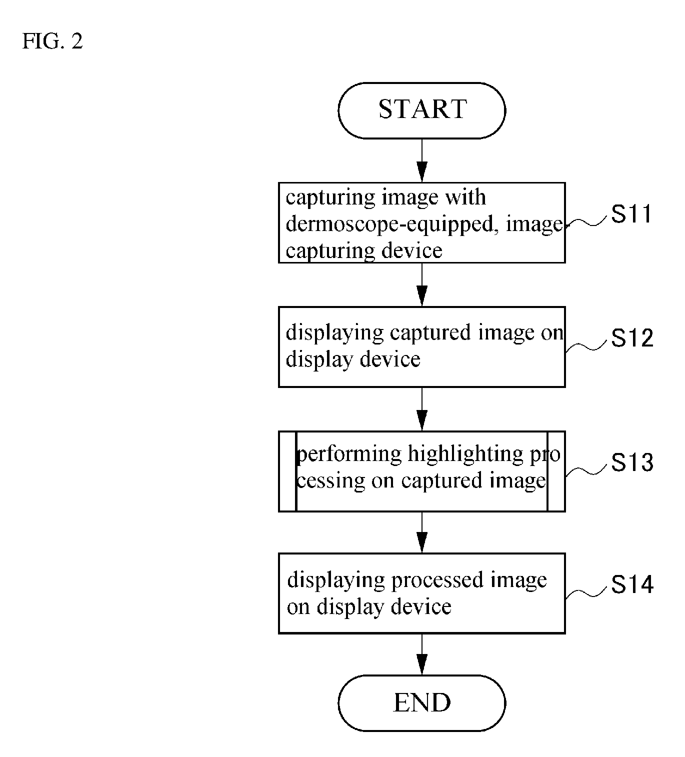
[0053]FIG. 8 is block diagram showing a configuration of Application Example 1 employing the diagnostic apparatus 100 in accordance of the embodiment. In the afore-mentioned diagnostic apparatus 100, the resultant image that is obtained by performing the edge preserving smoothing filter processing on the input image is the base image; the resultant image that is obtained by subtracting the base image from the input image is the detail image; and the reconstruction is performed such that the gain of the base image is decreased and the gain of the detail image is increased. Due to the above configuration, the image in which the gradient inversion as well as the halo phenomenon occurring in the steep edge region of the image are suppressed can be generated. In the following Application Example 1, the edge preserving smoothing filter processed image is further subject to processing to generate a highlighted image, thereby further improving the accuracy of diagnosis.

[0054]Referring to FI...

application example 2

[0074]In accordance with the afore-mentioned Application Example 1, the brightness component of the captured image is separated into the base image and the detail image; the base image is compressed more brightly than the center value or is subjected to the sharpness filter processing; and the brightness is restored from the highlighted base image and the detail image; and the highlighted image is generated using the color information component.

[0075]However, the same effect as Application Example 1 can be obtained by separating the brightness component into the base image and the detail image; performing the highlighting processing on the detail image in accordance with the likelihood of an object to be diagnosed; restoring the brightness from the base image and the highlighted detail image; and generating the highlighted image using the color information component. Application Example 2 is hereinafter described in detail with reference to FIGS. 8-10.

[0076]In Application Example 2,...


PUM


Abstract

The invention provides a method of processing an image in a diagnostic apparatus of diagnosing a cutaneous lesion using a cutaneous image, comprising the steps of: (a) obtaining a first detail image made by performing a first component separation filter on a brightness component of the cutaneous image; (b) obtaining a second detail image made by performing a second component separation filter on the brightness component of the cutaneous image, the second component separation filter having properties different from those of the first component separation filter; (c) generating a third detail image based on the first detail image and the second detail image; (d) newly generating a third base image based on the third detail image; and (e) combining the third detail image with the third base image to restore a brightness component and generate a highlighted image.

Description

CROSS-REFERENCE TO RELATED APPLICATIONS[0001]This application claims priority from Japanese Patent Application No. 2015-054328 filed on Mar. 18, 2015, which is incorporated herein by reference.TECHNICAL FIELD[0002]The present invention relates to an image processing method to diagnose a cutaneous lesion, a diagnostic apparatus used for the same method, and a medium storing program associated with the same method.BACKGROUND ART[0003]Generally, visual inspection is necessarily performed to diagnose a cutaneous lesion, thereby obtaining an amount of information. However, not only discrimination between a mole and a spot but also discrimination between a benign tumor and a malignant tumor are substantially difficult with a naked eye inspection and even a magnifying glass inspection. For the reasons, dermoscopic inspection in which a dermoscope-equipped camera is used to capture an image of a disease has been conventionally performed.[0004]The dermascope is a noninvasive diagnostic devic...

Claims


Application Information

Patent Timeline
IPC IPC(8): G06T7/00G06T5/50A61B5/00G06T5/00
CPCG06T7/0012G06T7/0081G06T5/002G06T5/50G06T2207/30096G06T2207/10024G06T2207/10056G06T2207/30088A61B5/444G06T5/20G06T2207/20028G06T2207/20192G06T2207/20224G06T7/11G06T5/70
Inventor NAKAJIMA, MITSUYASU
Owner CASIO COMPUTER CO LTD
Features
  • R&D
  • Intellectual Property
  • Life Sciences
  • Materials
  • Tech Scout
  • Unparalleled Data Quality
  • Higher Quality Content
  • 60% Fewer Hallucinations
Learn More