
Tissue removal and closure device

a tissue and closure technology, applied in the field of surgical instruments, can solve the problems of reducing the service life of the patient,

Active Publication Date: 2016-09-15
AESCULAP AG
View PDF6 Cites 143 Cited by
  • Summary
  • Abstract
  • Description
  • Claims
  • Application Information

AI Technical Summary

Benefits of technology

The present patent describes surgical devices for separating a region of tissue from a tissue structure and for fastening the separated tissue. The devices include a shaft, a first jaw with a cutting track, a second jaw with a fastening track, and a cutting element moveable along the cutting track. The devices can be used to remove abnormal growths, cysts, tumors, or other abnormalities in a base region of tissue. The devices have features that allow for precise tissue removal and closure, and can be used in a variety of procedures.

Problems solved by technology

Otherwise, inadvertent removal of the base tissue can create additional trauma via an opening in the base tissue.
Moreover, repairing any opening created as a result of removal of suspect tissue can require complicated or time consuming stitching or suturing to close the opening in the base tissue of the organ.
There is also a risk of post-surgical complications if the physician does not properly close the opening.
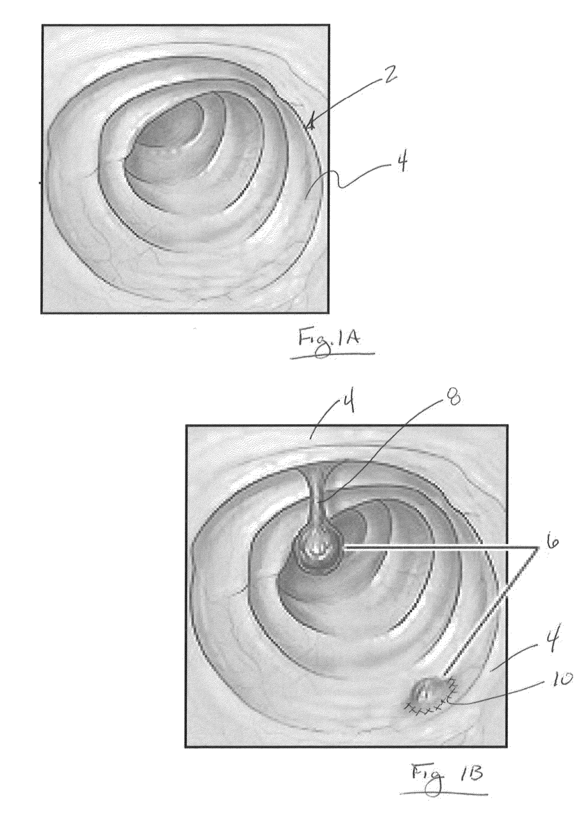
The risks associated with inadvertently creating an opening in an organ during removal of suspect tissue can present a dilemma for a physician that first observes the polyp (or other suspect tissue) but is hesitant to remove it if the physician believes that it will be difficult to maintain a clean margin (i.e., a region of relatively healthy tissue) at the site of the excision.
As shown, because the initial examinations are often performed via a minimally invasive procedure, the limited area or tortuosity within the organ 2 increases the difficulty of navigating and manipulating tools to the site for removal of tissue and / or closing of openings within the tissue 4.
In addition, the physician will often be hesitant to remove such a region of tissue if there is a risk that the procedure creates an opening in the wall of an organ where such an opening could lead to further complications to the patient.

Method used


Image

  • Tissue removal and closure device
  • Tissue removal and closure device
  • Tissue removal and closure device

Examples


Embodiment Construction

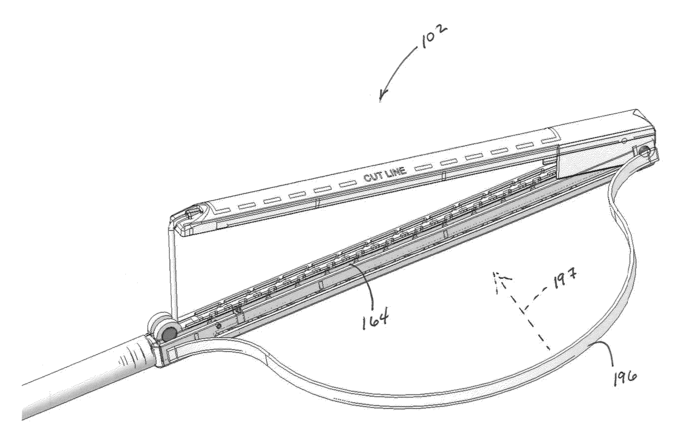
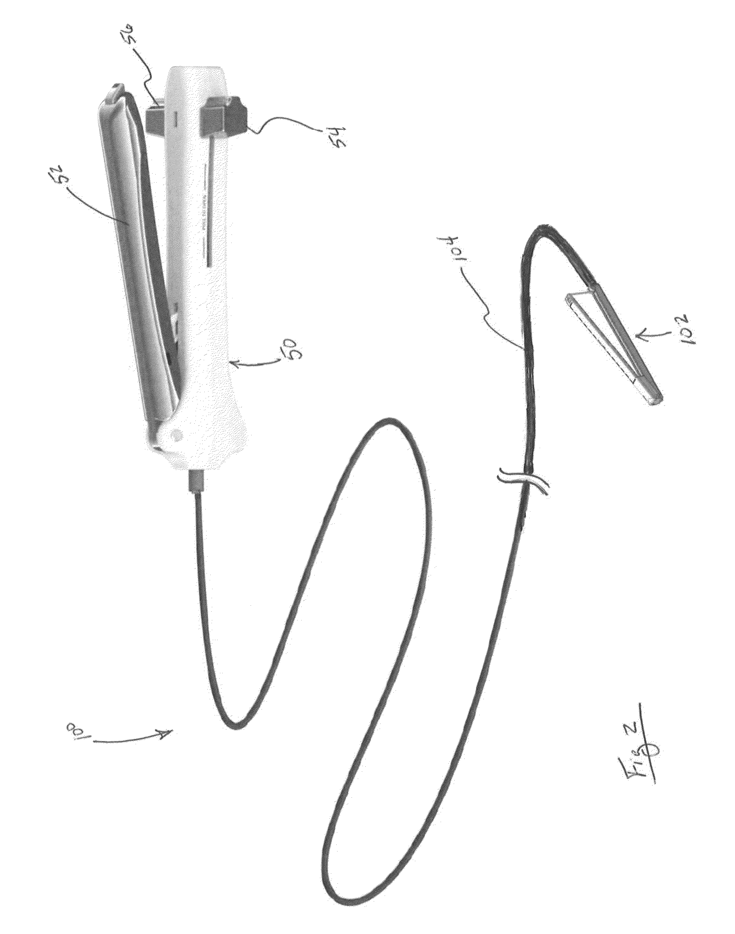
[0039]Methods and devices described herein provide for separating a suspect region of tissue from a base region of tissue and securing the base region of tissue that remains after the excision of the suspect tissue. Although the following description discusses the use of mechanical fasteners and a mechanical cutting instrument, any number of fastening modes and cutting modes can be employed in accordance with the disclosure herein as long as such modes do not conflict with the teachings disclosed herein. In addition, any number of combinations of aspects of various species, or combinations of the species themselves are within the scope of this disclosure. In addition, the methods, devices and systems described herein are well suited towards endoscopic procedures, but are not limited to such procedures. The disclosure can be combined with open procedures as well.

[0040]FIG. 2 illustrates an example of a surgical device 100 according to the present disclosure where the device 100 inclu...


PUM


Abstract

Methods and devices described herein facilitate improved treatment of body organs and relates to surgical instruments, useful in endoscopic, laparoscopic and / or open surgical procedures to effectively remove a suspect region of tissue such as a polyp, abnormal growth, cyst, tumor, lesion, or other abnormality from a base tissue structure.

Description

RELATED APPLICATIONS[0001]This application is a non-provisional of U.S. Patent Application No. 62 / 196,785 filed Jul. 24, 2015. This application is also a continuation-in-part of U.S. application Ser. No. 13 / 028,148 filed Feb. 15, 2011. The entirety of both applications are incorporated by reference in its entirety.FIELD OF THE INVENTIONS[0002]The present invention relates to surgical instruments, useful in endoscopic, laparoscopic and / or open surgical procedures to effectively remove a suspect region of tissue such as a polyp, abnormal growth, cyst, tumor, lesion, or other abnormality from a base tissue structure.BACKGROUND[0003]Minimally invasive surgical procedures are becoming more common as surgeons use a variety of techniques to operate with reduced trauma to the body than with open surgeries. In general, minimally invasive surgical procedures are safer than open surgeries and allow the patient to recover faster and heal with less pain and scarring. Minimally invasive surgical ...

Claims


Application Information

Patent Timeline
Patent Type & Authority Applications(United States)
IPC IPC(8): A61B17/072A61B17/32
CPCA61B17/072A61B17/32A61B2017/07271A61B2017/07285A61B2017/320052A61B17/07207A61B2017/00115A61B2017/07214A61B2017/07228A61B2017/07278A61B2017/2927A61B2017/2937A61B2090/0807
Inventor HAUSEN, BERRNARD A.DONOHOE, BRENDAN M.
Owner AESCULAP AG
Learn More