
Composition for regeneration of cartilage

a cartilage and collagen technology, applied in the direction of drugs, peptide/protein ingredients, prosthesis, etc., can solve the problems of lack of vascular and neural networks, lack of cartilage defect lesion repair ability, etc., to reduce the burden on patients

Inactive Publication Date: 2016-03-10
MOCHIDA PHARM CO LTD +1
View PDF3 Cites 1 Cited by
  • Summary
  • Abstract
  • Description
  • Claims
  • Application Information

AI Technical Summary

Benefits of technology

[0017]Under the above-described circumstances, there has been a need in the cartilage regeneration medicine for a composition for cartilage regeneration, which can reduce the burden on patients such as cell harvest, arthrotomy and the like, which can easily be applied to a cartilage injury lesion, and which can exert a cartilage regeneration effect in vivo. In particular, a novel composition for cartilage regeneration has been expected that can produce a good hyaline cartilage regeneration effect that resembles that of normal cartilage without using graft cells.
[0018]In order to solve the above-described problems, the present inventors have gone through intensive studies and found that good cartilage regeneration can be induced at a cartilage injury lesion without using graft cells by applying to the cartilage injury lesion a composition for cartilage regeneration wherein (a) a monovalent metal salt of a low endotoxin alginic acid and (b) SDF-1 are used in combination, thereby accomplishing the present invention.
[0021]The present invention provides a composition for cartilage regeneration in cartilage regeneration medicine, which can reduce the burden on patients such as cell harvest, arthrotomy and the like, which can easily be applied to a cartilage injury lesion, and which can exert cartilage regeneration effect in vivo. A composition for cartilage regeneration according to a preferred embodiment of the present invention is capable of inducing hyaline cartilage regeneration that resembles that of normal cartilage without using graft cells.

Problems solved by technology

Articular cartilage, however, lacks vascular and neural networks.
Accordingly, it has very little self-repairing ability and thus a cartilage defect lesion is not adequately repaired particularly when a large defect lesion is formed in the cartilage.
Therefore, a cartilage defect lesion leads to pain in the joint and loss of joint function, which often result in development of osteoarthritis.
In addition, an early stage of osteoarthritis, where abrasion of the surface of articular cartilage starts due to aging or excessive joint usage, may lead to cartilage defect over a large region as a result of progression of the symptoms.
Thus, since articular cartilage is inadequate in self-repairing ability, surgical procedures for treating cartilage lesion, for example, autologous osteochondral implantation (mosaicplasty), a procedure of perforating with a pick (microfracture), drilling, a procedure of shaving subchondral bone with a burr (abrasion procedure) and resection of injured cartilage (debridement) are required.
These techniques, however, have limitation for a widespread cartilage defect and cartilage regenerated by these procedures is fibrous cartilage that has different mechanical property from that of hyaline cartilage.
Recent clinical examinations, however, also report that ACI technique did not show significantly superior results over other surgeries with respect to repair of an articular cartilage defect.
One reason is the technical difficulties in anchoring cells and a scaffold to the joint defect lesion and covering them with a periosteum patch.
Moreover, several complicated issues related to the periosteum patch including thickening, defect and intra-articular adhesion of periosteum have also been reported.
The other reason is the limitation to the use of chondrocytes.
Another problem is that harvest of cartilage from a non-weight-bearing site of the affected joint required by the ACI technique leaves the problem of the donor site that has been harvested of chondrocytes to remain problematic.
It is, however, difficult to acquire sufficient cartilage regeneration effect with polymer alone, and polymer is usually used in combination with cultured chondrocytes and mesenchymal stem cells.
Generally, in cartilage regeneration medicine, it is difficult to predict the in vivo effect of implantation therapy only by in vitro cartilage regeneration tests or cell migration tests.
It is difficult to confirm such performances other than in vivo.

Method used


Image

  • Composition for regeneration of cartilage
  • Composition for regeneration of cartilage
  • Composition for regeneration of cartilage

Examples


example 1

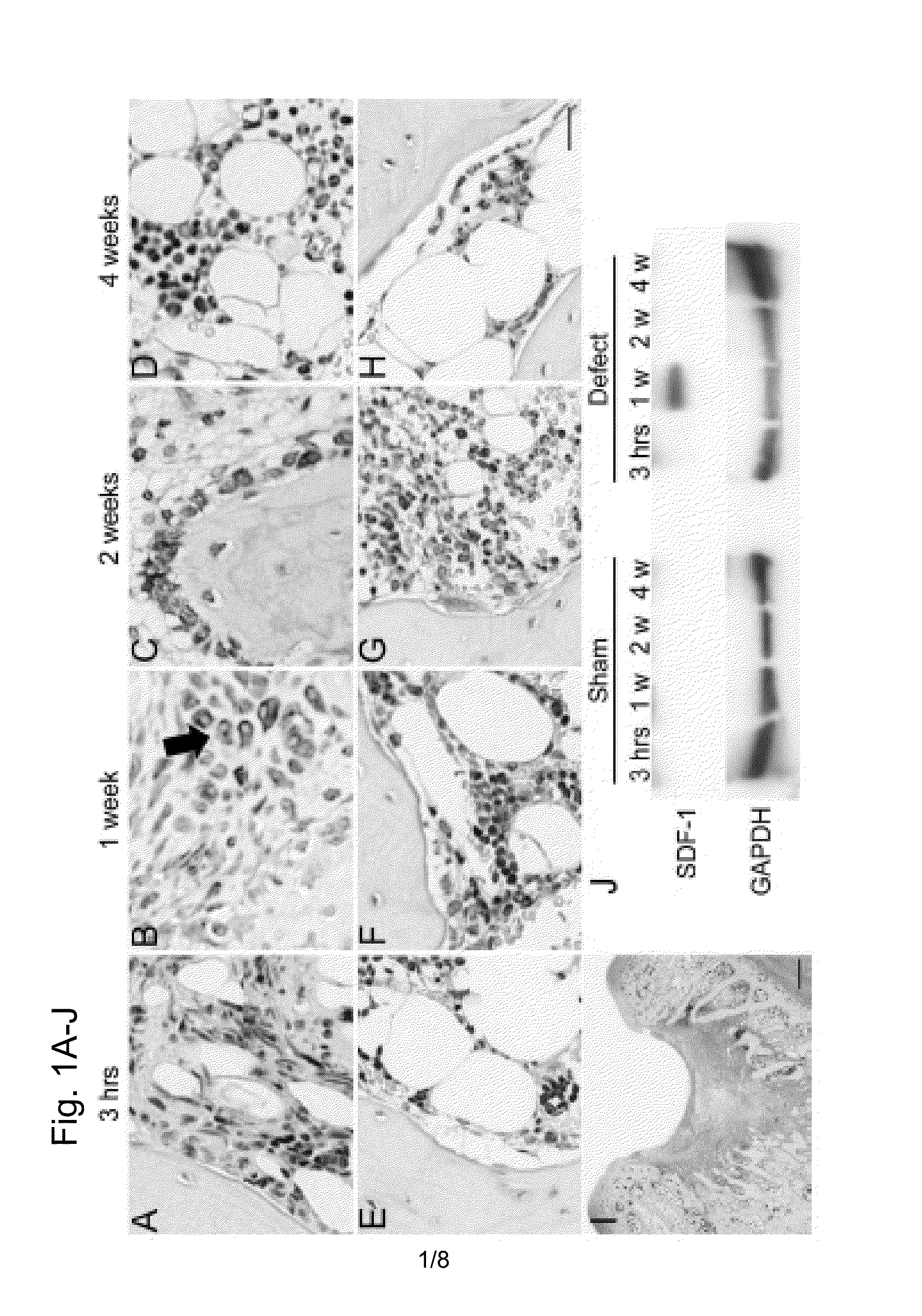
Expression of SDF-1 Protein in Osteochondral Defect Models

[0119]An osteochondral defect model of a rabbit knee joint was used to examine change in the expression of SDF-1 protein at a injury site with time by immunohistostaining.

[0120]15-week-old female Japanese white rabbits (weight 2.6 to 2.9 kg) were used to prepare osteochondral defect models. Anesthesia was performed by intravenous administration of 0.05 mg / kg pentobarbital and inhalation anesthesia of isoflurane. An antibiotic (penicillin G, Meiji Co., Ltd.) was intramuscularly administered. After incision in the skin of about 2 cm, patella was turned over by medial parapatellar approach to expand the patellar surface of the femur. A power drill (Rexon, Kawasaki) was used to create full-thickness osteochondral defects (diameter of 4.5 mm and depth of 3 mm) that reach the subchondral bones in the patellar surfaces of femurs of both knees. A sham surgery group (control) received the same surgery except the creation of the full-t...

example 2

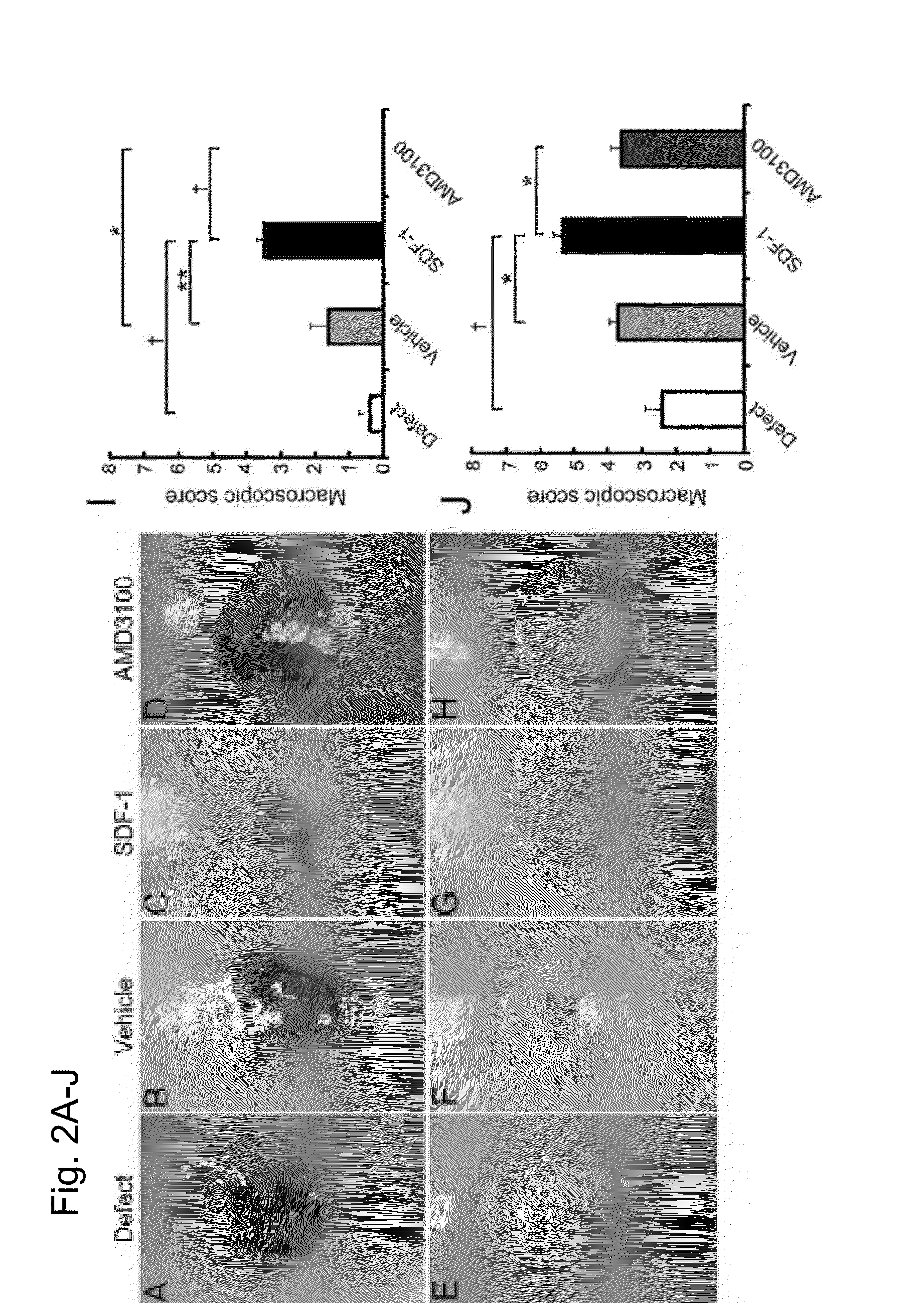
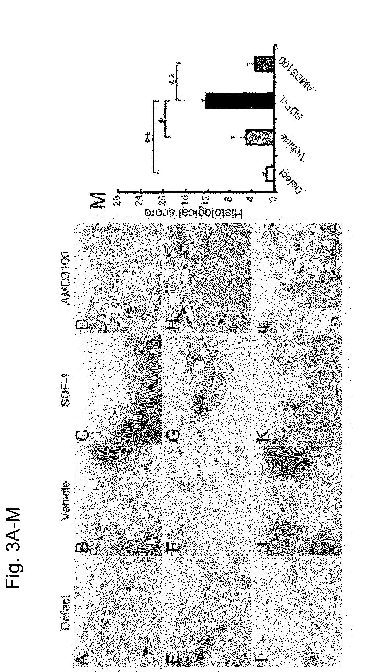
Application of Alginic Acid Gel to Rabbit Osteochondral Defect Models

[0125](2-1) Alginic Acid

[0126]A low endotoxin sodium alginate (Sea Matrix (sterilized), with a molecular weight of about 1,700,000 Da, distributed from Mochida International Ltd.) was used. 12.5 ml of deionized water sterilized by a filter procedure was added to 0.25 g of sodium alginate (lyophilized product) to obtain a 2% sodium alginate solution that is to be used in the experiment. The endotoxin level of the low endotoxin sodium alginate was 5.76 EU (endotoxin unit) / g. The endotoxin level of food grade (commercial grade) sodium alginate (Wako Pure Chemical Industries, Ltd., sodium alginate 500, 199-09961) is 75,950 EU / g, and thus sodium alginate with an extremely low endotoxin level is used in this experiment.

[0127](2-2) Preparation of Models

[0128]Rabbit osteochondral defect models were prepared by the procedure described in Example 1. The prepared full-thickness osteochondral defects were washed with physiolog...

example 3

Assessment of In Vivo Cell Homing

[0157]After applying the alginic acid+SDF-1 to the osteochondral defect lesion, quantitative assessment was conducted to see how the in vivo cell homing and migration to the injury lesion take place. An alginic acid group, an alginic acid+SDF-1 administration group and an alginic acid+AMD3100 administration group were prepared in the same method as the rabbit osteochondral defect models described in Example 2. Seven days after the surgery, the implanted alginic acid gels were collected and the numbers of cells in the gels were counted. The collected gel was immobilized with 4% phosphate buffered paraformaldehyde for 24 hours and embedded in paraffin to prepare a sample using a section from the center of the gel with a thickness of 5 μm. The samples were stained with H-E (hematoxylin and eosin).

[0158]A week after the surgery, more cells were found in the gel of alginic acid+SDF-1 administration group compared to other groups (FIG. 7C). From the quanti...


PUM

PropertyMeasurementUnit
concentrationaaaaaaaaaa

Abstract

A novel composition for regenerating a cartilage has been demanded, which can achieve a good effect of regenerating a hyaline cartilage that is a nearly normal cartilage without requiring the use of any transplanted cell. The present invention provides a composition for regenerating a cartilage, wherein (a) a monovalent metal salt of low endotoxin alginic acid and (b) SDF-1 are used in combination.

Description

CROSS-REFERENCE TO RELATED APPLICATIONS[0001]This application is a Continuation of U.S. application Ser. No. 14 / 239,663, which is the U.S. National Stage application of PCT / 2012 / 071749, filed Aug. 22, 2012, which claims priority from Japanese application JP 2011-181662, filed Aug. 23, 2011.TECHNICAL FIELD[0002]The present invention relates to a composition for cartilage regeneration and the like.BACKGROUND ART[0003]Articular cartilage is hyaline cartridge that is composed of a small number of cells, collagenous extracellular matrix, abundant proteoglycans and water. Since vascular and neural networks are present in the bone and the bone has self-repairing ability, even when a bone is fractured, the fractured site is often adequately repaired. Articular cartilage, however, lacks vascular and neural networks. Accordingly, it has very little self-repairing ability and thus a cartilage defect lesion is not adequately repaired particularly when a large defect lesion is formed in the cart...

Claims


Application Information

Patent Timeline
Patent Type & Authority Applications(United States)
IPC IPC(8): A61K38/19A61K31/734
CPCA61K31/734A61K38/195A61L27/20A61L27/54A61L2300/252A61L2430/06A61L2300/414C08L5/04
Inventor IWASAKI, NORIMASASUKEGAWA, ATSUSHIMINAMI, AKIOOHZAWA, NOBUO
Owner MOCHIDA PHARM CO LTD
Learn More