
Cell differentiation assay method, cell isolation method, method for producing induced pluripotent stem cells, and method for producing differentiated cells

Active Publication Date: 2015-04-23
FUJIFILM CORP +1
View PDF0 Cites 5 Cited by
  • Summary
  • Abstract
  • Description
  • Claims
  • Application Information

AI Technical Summary

Benefits of technology

The present invention relates to a lectin called BC2LCN that can specifically detect a sugar chain on undifferentiated stem cells. This lectin can be labeled with a substance and reacted with test cells to evaluate their differentiation status. This allows for the simple and effective evaluation of cell fate. The lectin can also be used to separate cells in an undifferentiated state and cells that have advanced differentiation. This makes the lectin useful in regeneration medicine and biologics.

Problems solved by technology

As the clinical application of human pluripotent stem cells such as ES cells and iPS cells are started as just described above, a system to supply cells by securing quality and safety is not sufficiently developed.
This becomes a cause of bringing negative effects such as the decreased reliability of stem cell therapy and the occurrence of health hazards due to the therapy.
For example, although pluripotent stem cells are not directly used but used after differentiating them into desired cells for transplantation in a cell therapy, it has been pointed out that if a cell source having differentiated into desired cells is contaminated with undifferentiated cells, these undifferentiated stem cells become a cause of tumorgenesis.
However, it is not easy to stably obtain cells having quality suitable for transplantation; thus, it represents a very important challenge to establish a quality verification method for somatic stem cells and a stable culture method based thereon.

Method used


Image

  • Cell differentiation assay method, cell isolation method, method for producing induced pluripotent stem cells, and method for producing differentiated cells
  • Cell differentiation assay method, cell isolation method, method for producing induced pluripotent stem cells, and method for producing differentiated cells
  • Cell differentiation assay method, cell isolation method, method for producing induced pluripotent stem cells, and method for producing differentiated cells

Examples


example 1

Cell Staining of Human ES Cell

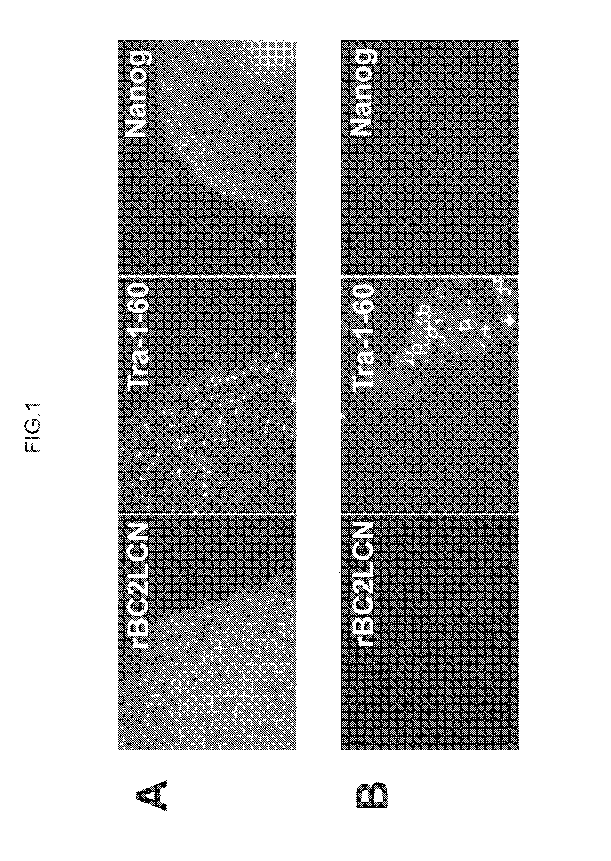
[0146]Human ES cells (KhES-1 strain) used in this Example were obtained from Institute for Frontier Medical Sciences, Kyoto University. These cells were cultured by the method of Suemori et al. (see Non Patent Literature 6). ES cell colonies were fixed with 4% paraformaldehyde and washed with PBS, to which rBC2LCN lectin fluorescently labeled (bound to Cy3) was then added for reaction at room temperature for 1 hour (A in FIG. 1). As a target for comparison, the stained image of the ES cell colonies is shown which was reacted with Tra-1-60 antibody or anti-Nanog antibody specifically recognizing undifferentiated ES cells or iPS cells and then further reacted with anti-mouse IgM-Alexa 488 or anti-rabbit IgG-Alexa 594 as a secondary antibody (A in FIG. 1). The fluorescence-labeled rBC2LCN lectin strongly stained the ES cells as did Tra-1-60 antibody or anti-Nanog antibody. Because the above experiment was performed without crushing cells, it is noted that ...

example 2

Staining of Human iPS Cell

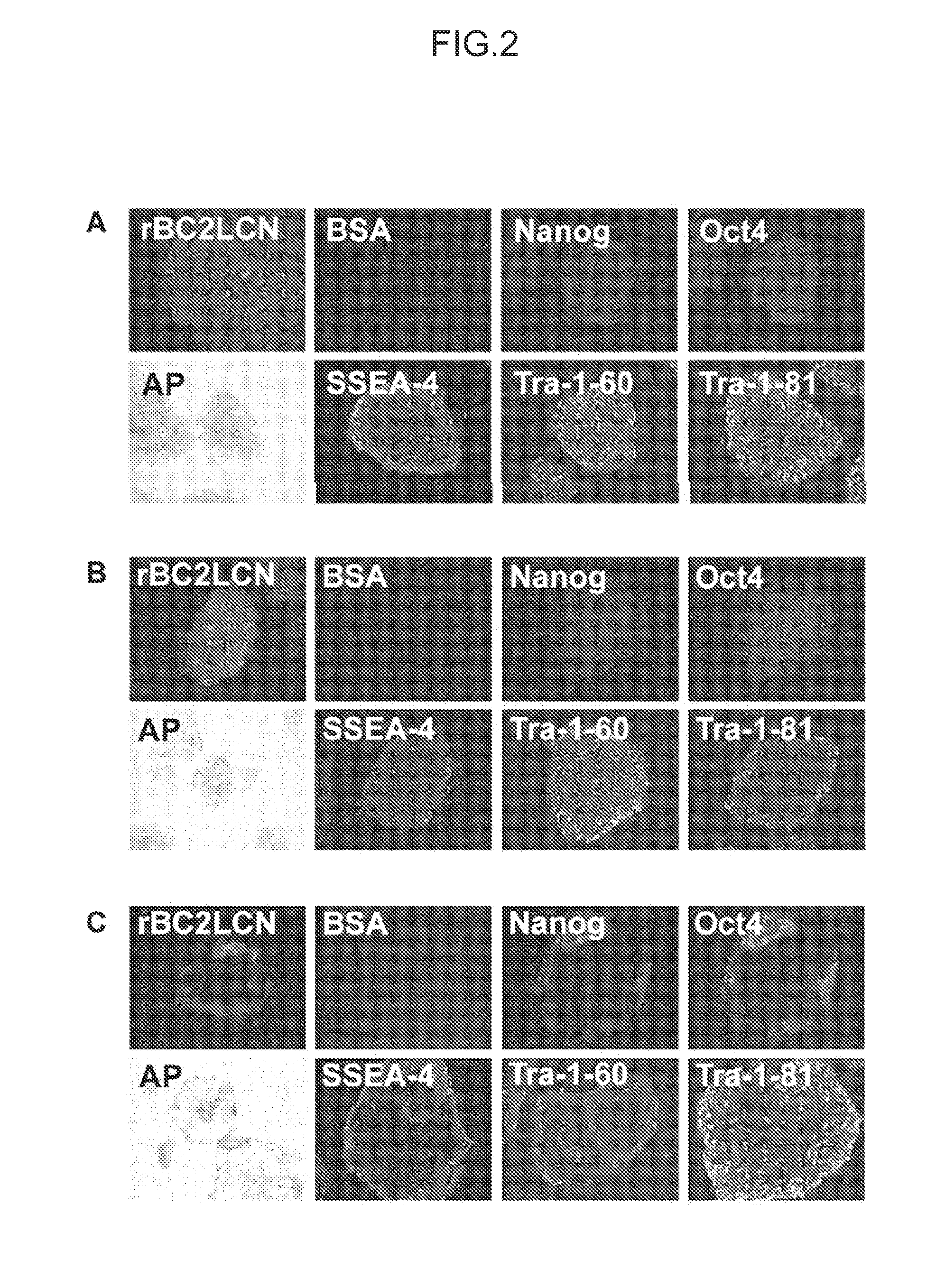
[0149]iPS cells (253G1 strain) used in this Example were obtained from the Riken BioResource Center. Cells were cultured by the method of Tateno et al. (see Non Patent Literature 1). Cells were fixed with 4% paraformaldehyde and washed with PBS, to which rBC2LCN lectin fluorescently labeled (bound to Cy3) was then added for reaction at room temperature for 1 hour (FIG. 2). Fluorescence-labeled BSA was used as a negative control (FIG. 2). As a target for comparison, staining with alkaline phosphatase as a marker for undifferentiated ES cells or iPS cells or staining with an antibody (anti-Nanog antibody, anti-Oct3 / 4 antibody, SSEA-4 antibody, Tra-1-60 antibody, or Tra-1-81 antibody) was performed (FIG. 2). It was shown that fluorescence-labeled BC2LCN lectin strongly stained undifferentiated iPS cells and strongly detected iPS cells to the same extent as or more extent than staining with AP as a known undifferentiation marker and various antibody staining (A...

example 3

Flow Cytometry of iPS Cell Using Fluorescence-Labeled BC2LCN Lectin

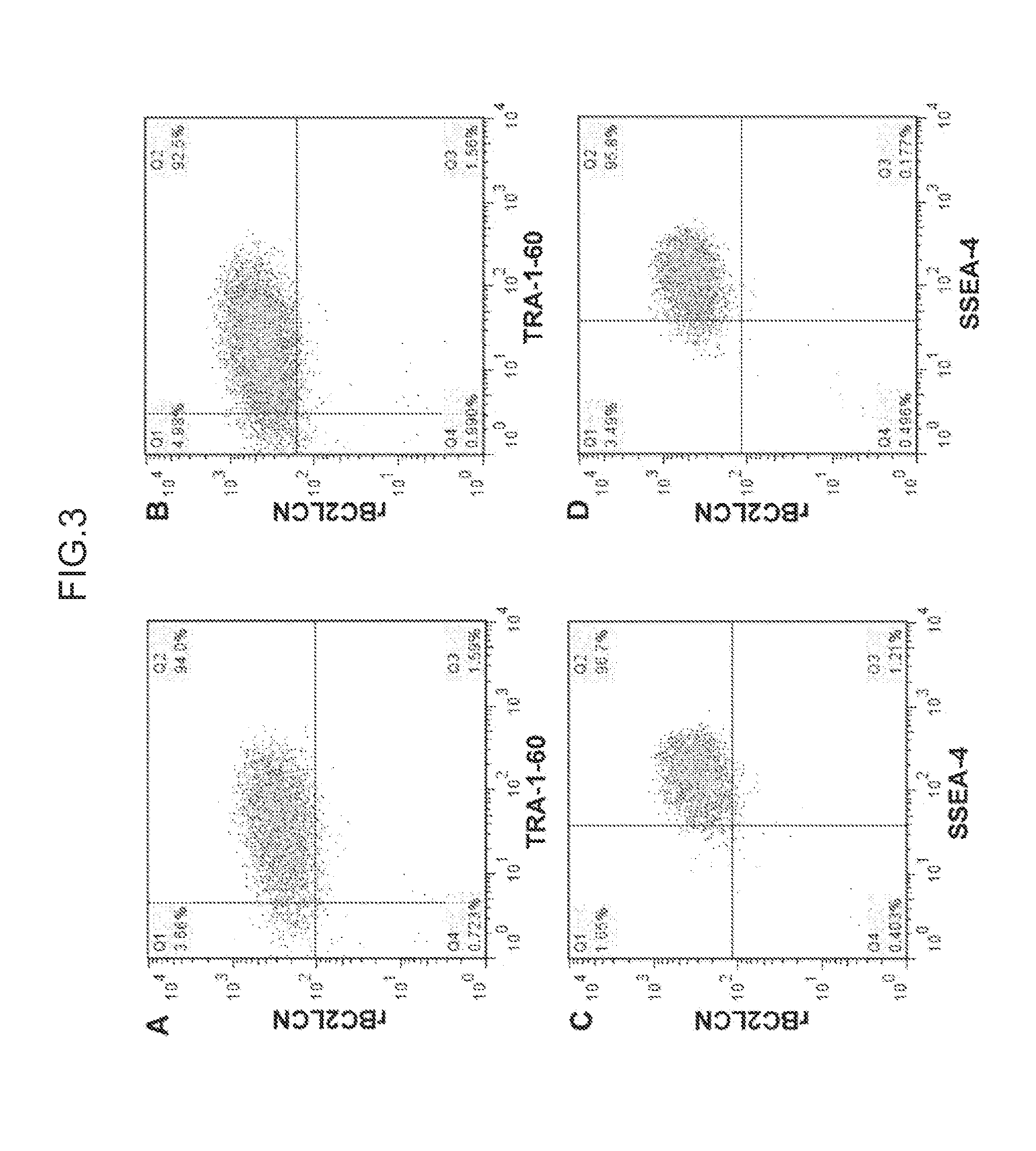
[0152]iPS cells (253G1 strain) prepared in the same way as in Example 2 were dissociated by enzyme treatment, reacted with fluorescence-labeled (HiLyte Fluor 647-bound) rBC2LCN lectin and a known undifferentiation detection antibody (Tra-1-60 antibody or SSEA-4 antibody), and subjected to flow cytometry analysis using a FACS apparatus (FIG. 3).

[0153]As a result, 92.5 to 94.0% of all cells simultaneously bound to Tra-1-60 antibody and rBC2LCN lectin (A and B in FIG. 3). In addition, 95.8 to 96.7% of all cells simultaneously bound to SSEA-4 antibody and rBC2LCN lectin (C and D in FIG. 3). These results demonstrate that BC2LCN lectin can effectively sort iPS cells maintaining undifferentiation.

[0154]No difference was observed in the percentage of cells simultaneously binding to rBC2LCN and Tra-1-60 antibody or SSEA-4 antibody between a case where cells were reacted first with rBC2LCN lectin and later with Tra-1-60 antib...


PUM


Abstract

Provided are a method for accurately evaluating the differentiation status of stem cells by selectively staining only stem cells in an undifferentiated state, and a method for positively isolating only stem cells in an undifferentiated state. Specifically provided is a method for determining differentiation of a cell comprising a step of contacting a test cell with a probe comprising protein (A) or (B) below and a step of detecting the presence of binding of the probe to the test cell. The method for determining differentiation of a cell is capable of detecting the presence or absence of an undifferentiated stem cell in test cells by using a probe that specifically reacts with undifferentiated stem cells and detecting the presence of bonding to the test cell. (A) A protein comprising an amino acid sequence shown in SEQ ID NO: 1 and recognizing a sugar chain structure of “Fucα1-2Galβ1-3GlcNAc” and / or “Fucα1-2Galβ1-3GalNAc;” and (B) a protein comprising an amino acid sequence showing 80% or more similarity to the amino acid sequence shown in SEQ ID NO: 1 and recognizing a sugar chain structure of “Fucα1-2Galβ1-3GlcNAc” and / or “Fucα1-2Galβ1-3GalNAc.”

Description

TECHNICAL FIELD[0001]The present invention relates to a method for determining differentiation of a cell, a cell separation method, a method for producing induced pluripotent stem cells, and a method for producing differentiated cells. More specifically, the present invention relates to a method for determining the differentiation status of test cells by using a protein recognizing a sugar chain structure specific for undifferentiated cells, and the like.BACKGROUND ART[0002]Pluripotent stem cells have attracted attention because of having the property of being capable of differentiating into various cells constituting the body and the property of being capable of maintaining their characteristics being undifferentiated, and are not only applied to drug discovery screening and elucidation of disease mechanisms but also under world-wide study as a material for regenerative medicine.[0003]The world's first phase 1 clinical trial using human ES cells started against acute spinal-cord in...

Claims


Application Information

Patent Timeline
IPC IPC(8): G01N33/569
CPCG01N33/56966
Inventor TATENO, HIROAKIHIRABAYASHI, JUNASASHIMA, MAKOTOITO, YUZURUONUMA, YASUKOWARASHINA, MASAKIFUKUDA, MASAKAZU
Owner FUJIFILM CORP
Learn More