Active colonoscopy training model and method of using the same
a colonoscopy and training model technology, applied in the field of colonoscopy training systems and methods, can solve the problems of difficult cover, further complicating the procedure, and reducing the visual feedback of the trainee, so as to achieve the effect of reducing the discomfort of each patient and reducing the difficulty of training
- Summary
- Abstract
- Description
- Claims
- Application Information
AI Technical Summary
Benefits of technology
Problems solved by technology
Method used
Image


Examples
example 1
Force Measurements
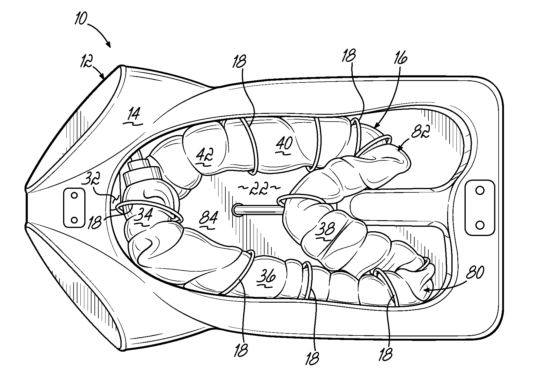
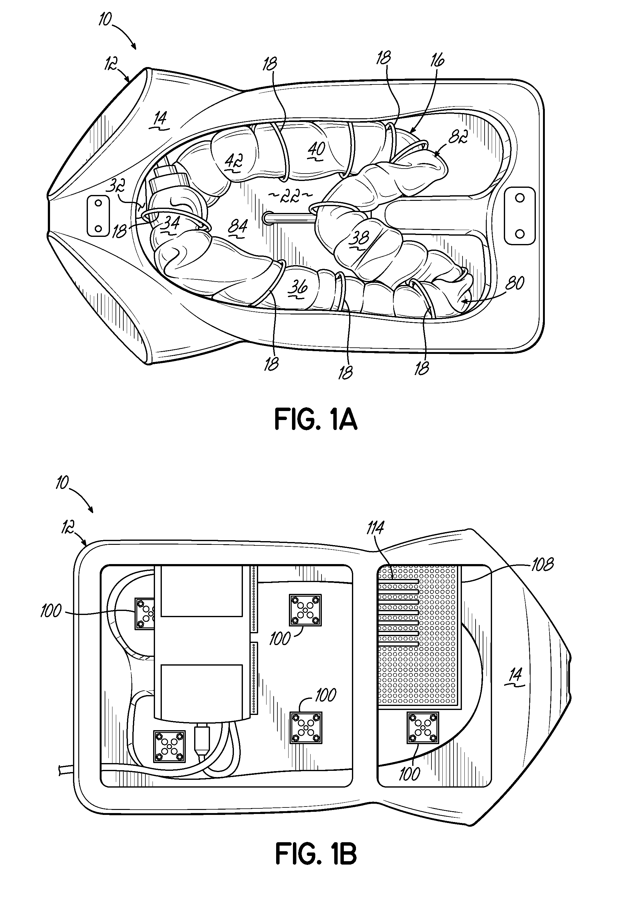
[0050]FIGS. 9A-9J and 10 illustrate the readouts of the forces measured during a complete colonoscopy training session with the ACTM 10 of FIGS. 1A-1B and the colonoscope 60 of FIG. 1C. Ten load cells 100 were arranged such that load cells L1, L2, and L3 were positioned along the sigmoid colon 34; load cells L4, L5, and L6 were positioned along the descending colon 36; load cell L7 was positioned at the splenic flexure 80; load cell L8 was positioned in the middle of the transverse colon 38; load cell L9 was positioned at the hepatic flexure 82; and load cell L10 was positioned in the middle of the ascending colon 40. During the colonoscopy training session, the colonoscopist performed a normal colonoscopy procedure and examined the inside of the colorectal tube 16 (even though the mucosa did not display any bowel diseases).
[0051]Table 1, below, shows the mean and maximum force detected by each load cell 100. For example, L1 detected the largest maximum over-all fo...
example 2
Localization of the Distal End
[0061]FIG. 11 illustrates the readouts of the photocells 104 of FIGS. 6A-6B during a complete colonoscopy training session with the ACTM of FIG. 1A and colonoscope 60 of FIG. 1C. Twenty-four photocells 104 were used with eight photocells 104 connected to each of Port 1, Port 2, and Port 0. The voltage data acquired by the data acquisition system 108, which were digital decimal values, were converted to 8 bit binary numbers and plotted against corresponding time in seconds and as shown in FIG. 11. The same information is presented quantitatively, below, in Table 2.
[0062]The detected voltage values at Port 1, to which P1-P8 are connected, indicate that the colonoscope 60 required about 75 sec to pass through the rectum 32 and the sigmoid colon 34. It should also be noted that Port 0, to which P9-P16 are connected, did not detect a voltage value until about 116 sec (1.9 min) into the procedure, which indicates that the distal end 70 of the colonoscope shaf...
example 3
Pressure Test
[0068]FIG. 12A and FIG. 12B illustrate the change in pressure within the ACTM 10 of FIG. 1A with respect to surgical time of a colonoscopy training session. During the colonoscopy training session, air was inserted through a colonoscope air duct until the colorectal tube 16 was fully expanded. After the air pressure reached peak value, the air flow was terminated and the colonic pressure decreased. The pressure measured at the cecum 42 started at about 0 mmHg and increased to 25 mmHg. The pressure was maintained at about 20 mmHg and showed a sudden increase at about 35 sec in the surgical time. While the anus unit (not shown) had been pressurized, some loss of pressure was expected due to the nature of the artificial organ. During a real colonoscopy procedure, some amount of the air inserted to the colon for opening the lumen may be initially lost through the anus, the anus unit (not shown) of the ACTM 10 demonstrated a continuous loss of air.
[0069]FIG. 12A and FIG. 12B...
PUM
 Abstract
Description
Claims
Application Information
 - R&D
- Intellectual Property
- Life Sciences
- Materials
- Tech Scout
- Unparalleled Data Quality
- Higher Quality Content
- 60% Fewer Hallucinations
Browse by: Latest US Patents, China's latest patents, Technical Efficacy Thesaurus, Application Domain, Technology Topic, Popular Technical Reports.
© 2025 PatSnap. All rights reserved.Legal|Privacy policy|Modern Slavery Act Transparency Statement|Sitemap|About US| Contact US: help@patsnap.com