System for ultrasound image guided procedure

Inactive Publication Date: 2014-09-11
EZONO
View PDF23 Cites 134 Cited by
  • Summary
  • Abstract
  • Description
  • Claims
  • Application Information

AI Technical Summary

Benefits of technology

The patent describes a system for detecting the position of a magnetic tool in the body using ultrasound images. This system has the advantages of using a standard magnetised tool and a freehand ultrasound probe. It also takes into account the imperfections of the model-based approach to improve accuracy. The system includes a look-up table that can be populated by direct measurements and interpolation of the measurements to reduce the time needed to measure the magnetic field. The system provides image information and detected position information to the clinician without substantial modification of the instruments used by the clinician and without needing substantial modification of the surgical procedures.

Problems solved by technology

In general it is very difficult for the user to determine the exact position of the tip of the medical tool and thus to be sure whether it is in the desired place, for example adjacent a nerve, or whether it has undesirably penetrated something else, for example a blood vessel.
However in view of the risks associated with exposure to electromagnetic radiation, it is not possible to provide a continuous guidance during insertion of the medical tool and so a series of snapshots are relied upon, which does not give optimal guidance.
However ultrasound imaging has two main challenges: firstly that the interpretation of ultrasound images is rather difficult, and secondly that needles do not show-up particularly reliably or visibly in the ultrasound image.
Further, even when the needle is in the imaging plane, because the echogenicity of standard needles is poor at high angles of incidence, the needle may not be particularly visible.
However these only help when the needle is well-aligned with the imaging plane.
Similarly techniques for image processing and ultrasound beam steering help only when the needle is well-aligned with the imaging plane and do not work well for angles of incidence greater than 45 degrees.
Various needle tracking technologies have been proposed based either on a needle guide fitted to an ultrasound probe, e.g. U.S. Pat. No. 6,690,159 or WO-A-2012 / 040077, or based on the transmission and reception of electromagnetic information, e.g. US-A-2007-027390) but have functional and accuracy limitations which means that the needle tip position is not exactly known in every clinical circumstance.
Further they often require the use of heavily modified or new equipment which is unwelcome to clinicians and to institutions with relatively rigid purchasing regimes.
Developing this level of skill and experience is time-consuming and difficult and as there is an anatomical variation from patient to patient, the procedures inevitably entail some risks.
In summary, although ultrasound guidance has improved some needling procedures, there are still significant difficulties and it cannot be used for many procedures.
This is a major barrier to its widespread use, particularly its use by practitioners who are not medical imaging specialists, such as anaesthetists, surgeons, pathologists, emergency physicians etc.
Imperfections compared to idealized model-based approaches introduce errors in position detection.

Method used


Image

  • System for ultrasound image guided procedure
  • System for ultrasound image guided procedure
  • System for ultrasound image guided procedure

Examples


Embodiment Construction

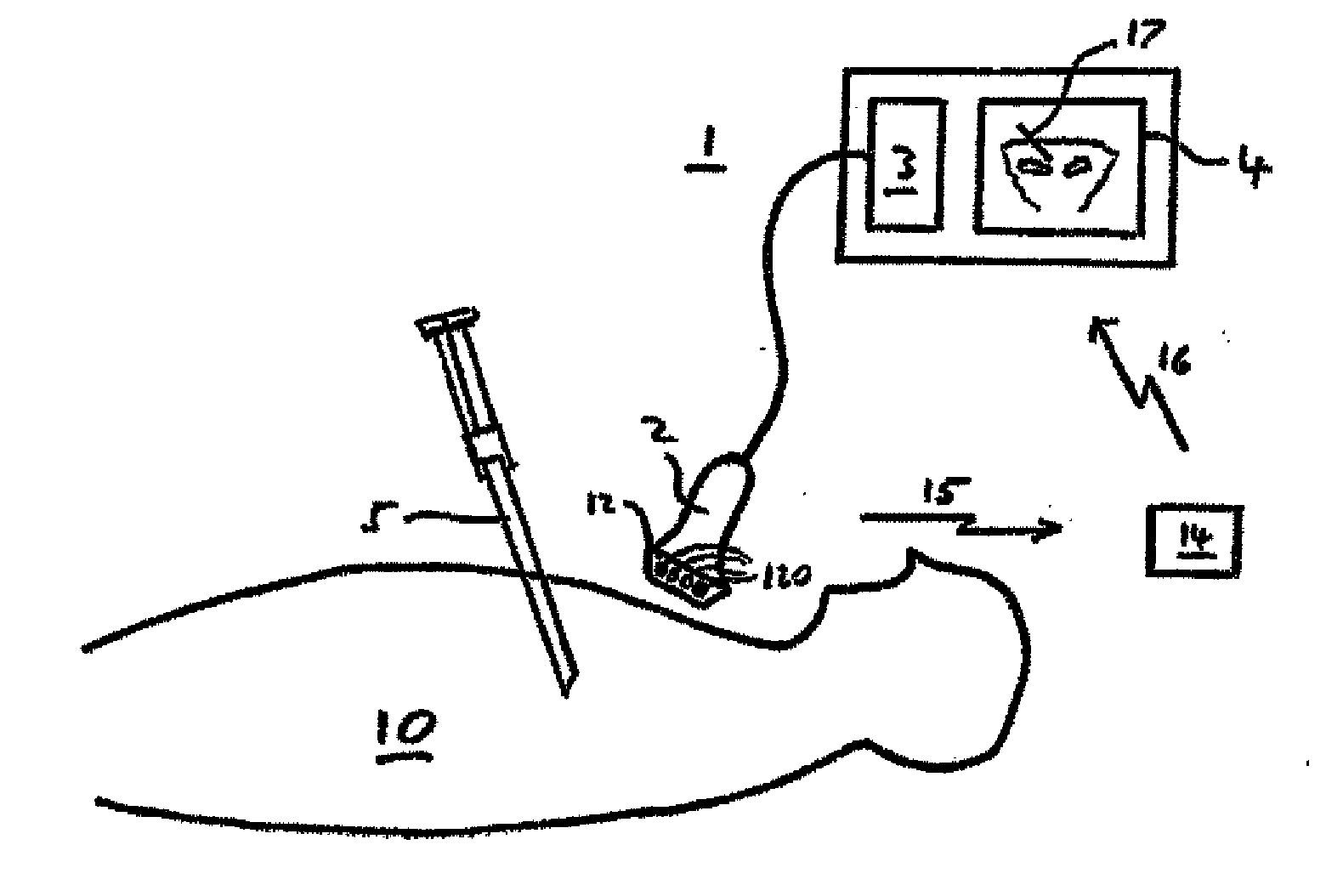
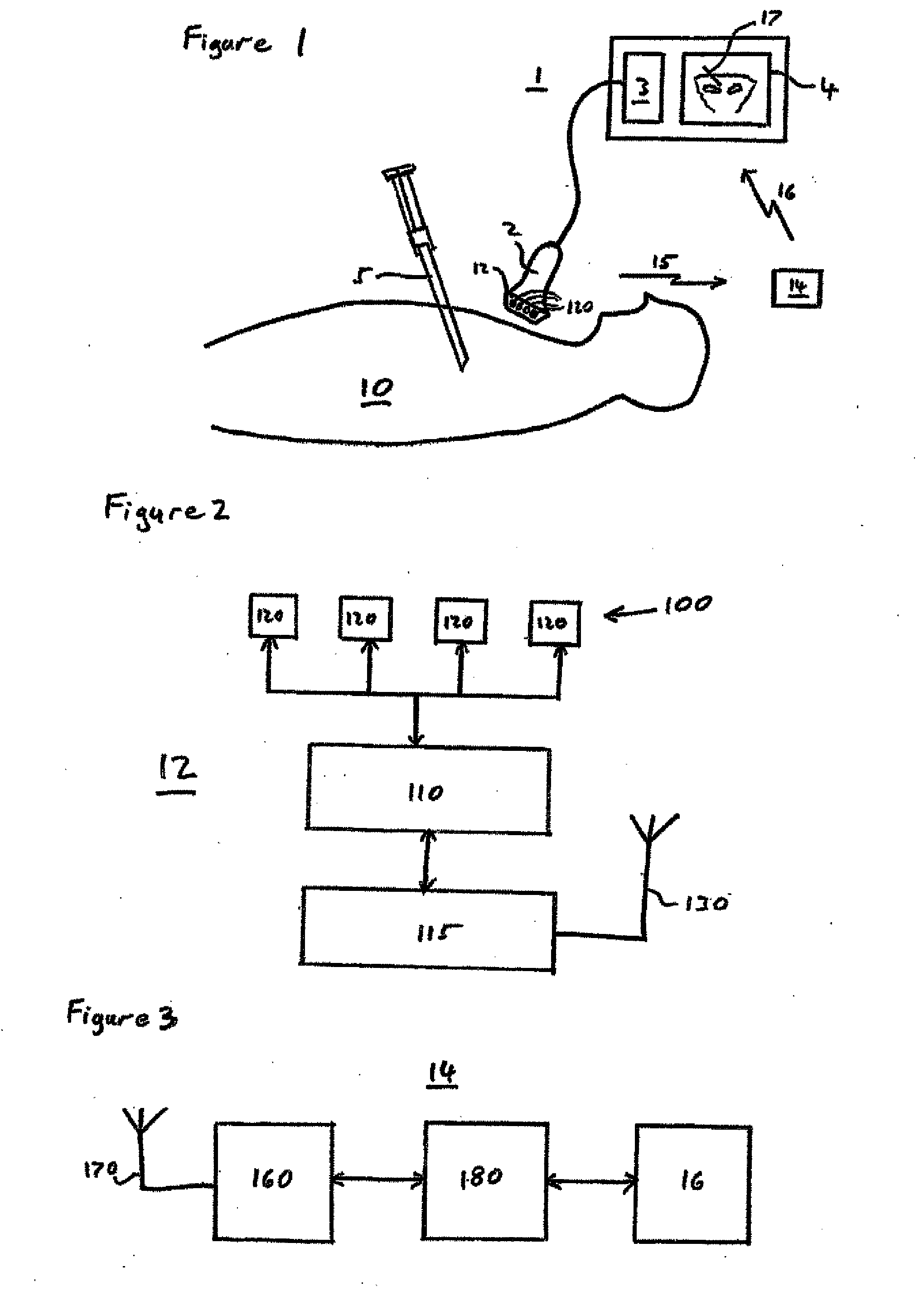
[0033]As shown in FIG. 1 the system in this embodiment of the invention comprises an ultrasound imaging system 1 including an ultrasound transducer 2, processor 3 and display 4. The system also comprises a tissue-penetrating medical tool 5 such as a needle, stylet, catheter or cannula.

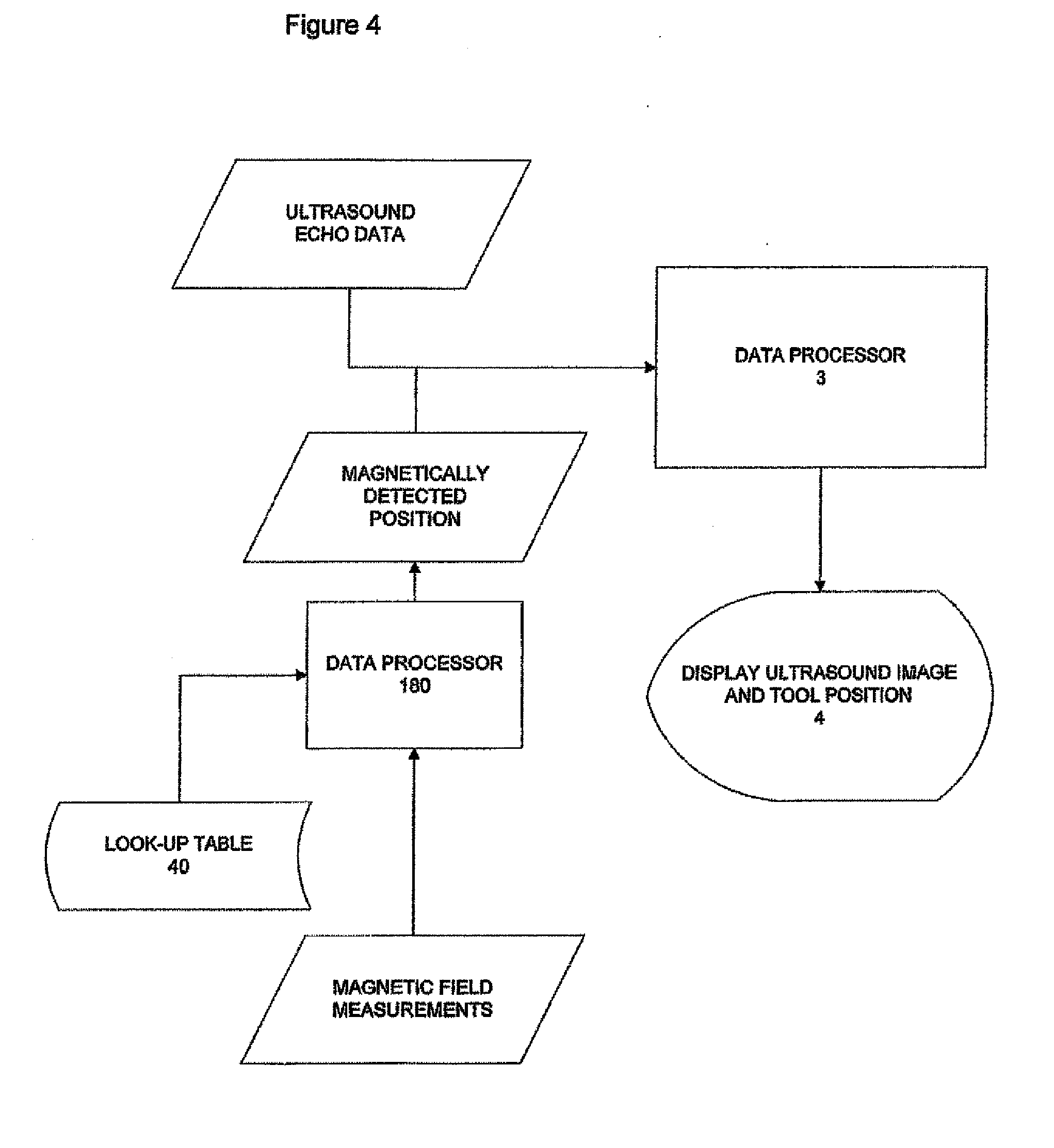
[0034]The invention uses magnetic position detection to track the tissue penetrating tool 5. Thus in this embodiment the tool 5 is magnetised and the ultrasound transducer 2 is provided with a magnetometric detector 12 comprising an array 100 of magnetometers 120. The detector 12 senses the magnetic field from the tool 5, together with the terrestrial magnetic field and any other background magnetic field, and the processor 3 is adapted to determine from the detected field the position and orientation of the tool 5 relative to the transducer 2. This magnetically detected position is then displayed on the display 4 together with the ultrasound image.

[0035]The ultrasound system 1 can be a standard two di...


PUM


Abstract

A system and method for ultrasound image guided surgical procedures such as needling or catheterisation, in which an ultrasound transducer is provided with a magnetometric detector for detecting the magnetic field emanating from a magnetised tissue-penetrating medical tool such as a needle or catheter. The detection of the magnetic field allows the position of the tool to be tracked magnetically and the position can be displayed on the ultrasound image. The position of the tool is determined from the magnetic field measurements by use of a look-up table of magnetic field values for the field emanating from the tissue-penetrating medical tool.

Description

TECHNICAL FIELD[0001]The present invention relates generally to the field of medical devices and in particular to a system for improving image guided procedures such as needle or catheterisation procedures.BACKGROUND AND OVERVIEW[0002]Unless explicitly indicated herein, the materials described in this section are not admitted to be prior art.[0003]There are numerous medical procedures that involve the insertion of a medical tool or instrument, such as a needle, cannula, catheter or stylet, into a subject's body, e.g. minimally-invasive surgical procedures, regional anaesthesia, detection of bio-electrical signals, electrical stimulation for diagnosis or treatment, vascular access, fine needle aspiration, musculoskeletal injections and so on. In such procedures it is generally necessary to guide the medical tool properly to the desired position in the subject's body and it can also be beneficial to monitor or track the medical tool position to ensure that it remains at the desired lo...

Claims


Application Information

Patent Timeline
IPC IPC(8): A61B8/00A61B5/06A61M25/01A61B5/00A61B17/34A61M25/09A61B5/05A61B8/08
CPCA61B8/4416A61B17/34A61B5/05A61B5/062A61M25/09A61B8/0841A61B8/52A61M25/0108A61M25/0127A61B5/72A61B2017/3413A61B8/4245A61B2034/2051A61B2090/378
Inventor DUNBAR, ALLANSOBRINO, ELISEO
Owner EZONO
Why Eureka
Try Eureka