Systems and methods for electrical stimulation of sphenopalatine ganglion and other branches of cranial nerves
a technology of cranial nerves and electrical stimulation, which is applied in the field of systems and methods for electrical stimulation of sphenopalatine ganglion and other branches of cranial nerves, can solve the problems of nasal mucosa sloughing, difficult to reliably perform pterygopalatine fossa injection, and only a brief respite from pain
- Summary
- Abstract
- Description
- Claims
- Application Information
AI Technical Summary
Benefits of technology
Problems solved by technology
Method used
Image
Examples
first embodiment
[0052]In the first embodiment, the stimulator is placed atop the mucosa area shown by hash marks in FIG. 6A, and medical glue such as cyanoacrylates or even denture adhesives are used to secure the stimulator to the mucosal surface. A surgical net may be placed over the stimulator to help keep it in place. Alternatively, the stimulator is attached to the underlying tissue using sutures that are placed through loops that are attached to the stimulator. This first, essentially noninvasive, embodiment might in some cases be considered to be temporary. When used temporarily, it may be used to determine whether the electrical stimulation will be effective, before using a more invasive approach. A significant advantage of this method is that there is no risk of damaging the blood vessels and nervous tissue within the pterygopalatine (sphenopalatine) fossa, because the fossa is not breached. Another advantage is its relative technical simplicity.
[0053]In the second embodiment (submucosal a...
third embodiment
[0054]The third embodiment is like the second, except that the anchor is inserted more permanently into a rigid structure such as the pterygoid process (PP in FIG. 6B) or the vomer. An example of such an anchor is shown in FIG. 7D. The stimulator is then attached to the anchor outside of the pterygopalatine (sphenopalatine) fossa at the site of attachment shown in FIG. 7D. Thus, the stimulator in the first three embodiments lies mostly outside the fossa.
fourth embodiment
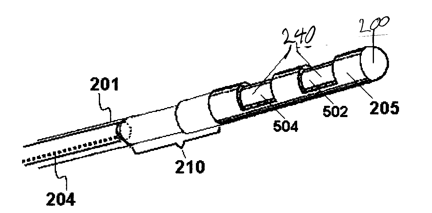
[0055]In the fourth embodiment, a needle is inserted through the mucosa by approximately 1 mm, into the pterygopalatine (sphenopalatine) fossa, at a site that is unlikely to encounter blood vessels or nerves. The site may have been selected by using high resolution MR imaging of the contents of the fossa, ultrasound imaging with a transducer at the tip of a catheter inserted through the nostril, or by the statistical likelihood of a safe insertion at some site. A lead blank (e.g., teflon coated flexible wire, possibly with a spatula-shaped end) is then inserted into the lumen of the needle. The lead blank is advanced along a path of least resistance, e.g., through fat tissue, but being deflected from the more solid nerve and blood vessel tissue as it advances. Thus, the path that the lead blank takes is not necessarily known in advance, but instead opens a safe wormhole within the fossa. The lead blank is then withdrawn from the needle, and a catheter-like stimulator is advanced int...
PUM
 Abstract
Description
Claims
Application Information
 - R&D
- Intellectual Property
- Life Sciences
- Materials
- Tech Scout
- Unparalleled Data Quality
- Higher Quality Content
- 60% Fewer Hallucinations
Browse by: Latest US Patents, China's latest patents, Technical Efficacy Thesaurus, Application Domain, Technology Topic, Popular Technical Reports.
© 2025 PatSnap. All rights reserved.Legal|Privacy policy|Modern Slavery Act Transparency Statement|Sitemap|About US| Contact US: help@patsnap.com


