System and Method for Real-Time Ultrasound Guided Prostate Needle Biopsy Based on Biomechanical Model of the Prostate from Magnetic Resonance Imaging Data

a biomechanical model and ultrasound technology, applied in the field of ultrasound guided prostate needle biopsies, can solve the problems of data for this study being subjected to a tedious visual inspection, low sensitivity of approximately 60% of the overall procedure results, and 25% positive predictive valu

Inactive Publication Date: 2013-12-05
SIEMENS MEDICAL SOLUTIONS USA INC
View PDF5 Cites 89 Cited by
  • Summary
  • Abstract
  • Description
  • Claims
  • Application Information

AI Technical Summary

Benefits of technology

The present invention is a method and system for using ultrasound to guide the insertion of a needle for prostate biopsy. The invention uses magnetic resonance imaging data to create a biomechanical model of the prostate and to help align the needle at the location of tumors. This is an automated process, important for clinical implementation. During the biopsy, the system maps the location of the tumors onto the ultrasound images in real-time, accounting for changes in the scanning plane and deformations of the prostate caused by the ultrasound probe. Overall, the invention improves the accuracy of needle insertion and reduces the likelihood of complications during prostate biopsy.

Problems solved by technology

The introduction of image guided biopsy using US has substantially increased the accuracy of biopsy, as compared with the very poor accuracy of performing blind biopsy, resulting in transrectal US guidance becoming the universally accepted methodology for prostate biopsy.
However, while transrectal US guided prostate biopsy is a clinically accepted methodology, the overall procedure results demonstrate a low sensitivity of approximately 60%, with only 25% positive predictive value.
The data for this study underwent a tedious visual inspection, which cannot be implemented as a clinical routine.

Method used


Image

  • System and Method for Real-Time Ultrasound Guided Prostate Needle Biopsy Based on Biomechanical Model of the Prostate from Magnetic Resonance Imaging Data
  • System and Method for Real-Time Ultrasound Guided Prostate Needle Biopsy Based on Biomechanical Model of the Prostate from Magnetic Resonance Imaging Data
  • System and Method for Real-Time Ultrasound Guided Prostate Needle Biopsy Based on Biomechanical Model of the Prostate from Magnetic Resonance Imaging Data

Examples


Embodiment Construction

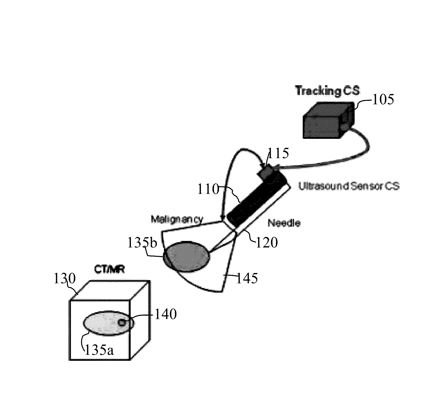
[0018]The present invention relates to a method and system for ultrasound (US) guided prostate needle biopsy based on a biomechanical model of the prostate from magnetic resonance (MR) imaging data. Embodiments of the present invention are described herein to give a visual understanding of the US guided prostate biopsy method. A digital image is often composed of digital representations of one or more objects (or shapes). The digital representation of an object is often described herein in terms of identifying and manipulating the objects. Such manipulations are virtual manipulations accomplished in the memory or other circuitry / hardware of a computer system. Accordingly, is to be understood that embodiments of the present invention may be performed within a computer system using data stored within the computer system.

[0019]Embodiments of the present invention provide increased sensitivity to prostate biopsy procedures to detect prostate cancer. Typically, a prostate biopsy id perfo...


PUM


Abstract

A method and system for real-time ultrasound guided prostate needle biopsy based on a biomechanical model of the prostate from 3D planning image data, such as magnetic resonance imaging (MRI) data, is disclosed. The prostate is segmented in the 3D ultrasound image. A reference patient-specific biomechanical model of the prostate extracted from planning image data is fused to a boundary of the segmented prostate in the 3D ultrasound image, resulting in a fused 3D biomechanical prostate model. In response to movement of an ultrasound probe to a new location, a current 2D ultrasound image is received. The fused 3D biomechanical prostate model is deformed based on the current 2D ultrasound image to match a current deformation of the prostate due to the movement of the ultrasound probe to the new location.

Description

[0001]This application claims the benefit of U.S. Provisional Application No. 61 / 653,552, filed May 31, 2012, the disclosure of which is herein incorporated by reference.BACKGROUND OF THE INVENTION[0002]The present invention relates to ultrasound guided prostate needle biopsies, and more particularly, to ultrasound guided prostate needle biopsies based on a biomechanical model of the prostate from magnetic resonance imaging data.[0003]Prostate cancer is typically diagnosed by using a transrectal ultrasound (US) guided biopsy, which is typically prescribed as a result of an elevated prostate-specific antigen (PSA) level or due to the detection of a palpable nodule during a digital rectal examination (DRE). The introduction of image guided biopsy using US has substantially increased the accuracy of biopsy, as compared with the very poor accuracy of performing blind biopsy, resulting in transrectal US guidance becoming the universally accepted methodology for prostate biopsy. However, ...

Claims


Application Information

Patent Timeline
IPC IPC(8): A61B8/08A61B10/02A61B5/055A61B8/13
CPCA61B8/0841A61B8/13A61B8/5261A61B10/0233A61B5/055A61B8/485A61B5/7425A61B8/14A61B8/4254A61B8/463A61B8/483A61B10/0241G06T2207/10136G06T2207/30081G06T7/33
Inventor KAMEN, ALIMANSI, TOMMASOGEORGESCU, BOGDANCOMANICIU, DORIN
Owner SIEMENS MEDICAL SOLUTIONS USA INC
Why Eureka
Try Eureka