
Endoscopic systems and methods for resection of tissue

a tissue resection and endoscopic technology, applied in the field of medical devices, can solve the problems of inadvertent cauterization or searing of healthy or non-target tissue, inability to precisely cut tissue, and inability to accurately detect the presence of a cutting instrument, so as to achieve enhanced tissue biopsy and faster use

Active Publication Date: 2013-06-20
COOK MEDICAL TECH LLC
View PDF7 Cites 15 Cited by
  • Summary
  • Abstract
  • Description
  • Claims
  • Application Information

AI Technical Summary

Benefits of technology

The present patent describes an apparatus and methods for resecting tissue using an endoscope. The apparatus includes a first tube with a working lumen and a second tube around it. A wire is attached to the second tube and can be rotated to incise tissue within the first tube's working lumen. The apparatus can provide a safer and faster way to resect desired tissue while minimizing damage to surrounding tissue. It can also improve tissue biopsy and reduce the number of "dry taps."

Problems solved by technology

However, when cutting instruments, such as needle knives, are used in the above-described manner, the result may yield an imprecise cutting of tissue.
In particular, a risk exists of improperly maneuvering the cutting instrument and therefore inadvertently cauterizing or searing healthy or non-target tissue.
Further, in these procedures, the extended cutting instrument is generally moved in a circular motion, which may increase the risk of incision to the unrelated tissue.
However, such biopsy needles occasionally result in “dry taps,” in which a column of tissue is pressed into the needle but not detached from surrounding tissue mass, thereby failing to collect the desired sample tissue.

Method used


Image

  • Endoscopic systems and methods for resection of tissue
  • Endoscopic systems and methods for resection of tissue
  • Endoscopic systems and methods for resection of tissue

Examples


Embodiment Construction

[0021]In the present application, the term “proximal” refers to a direction that is generally towards a physician during a medical procedure, while the term “distal” refers to a direction that is generally towards a target site within a patient's anatomy during a medical procedure.

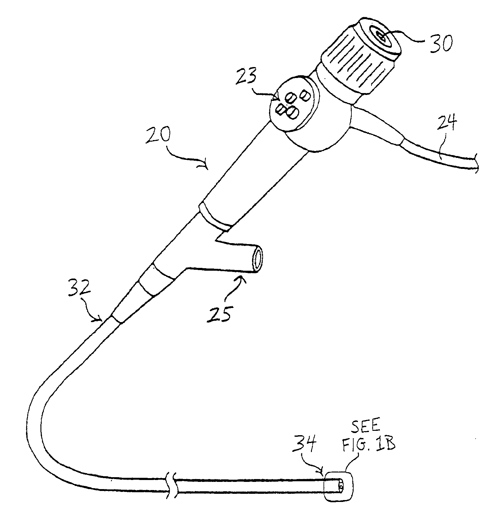
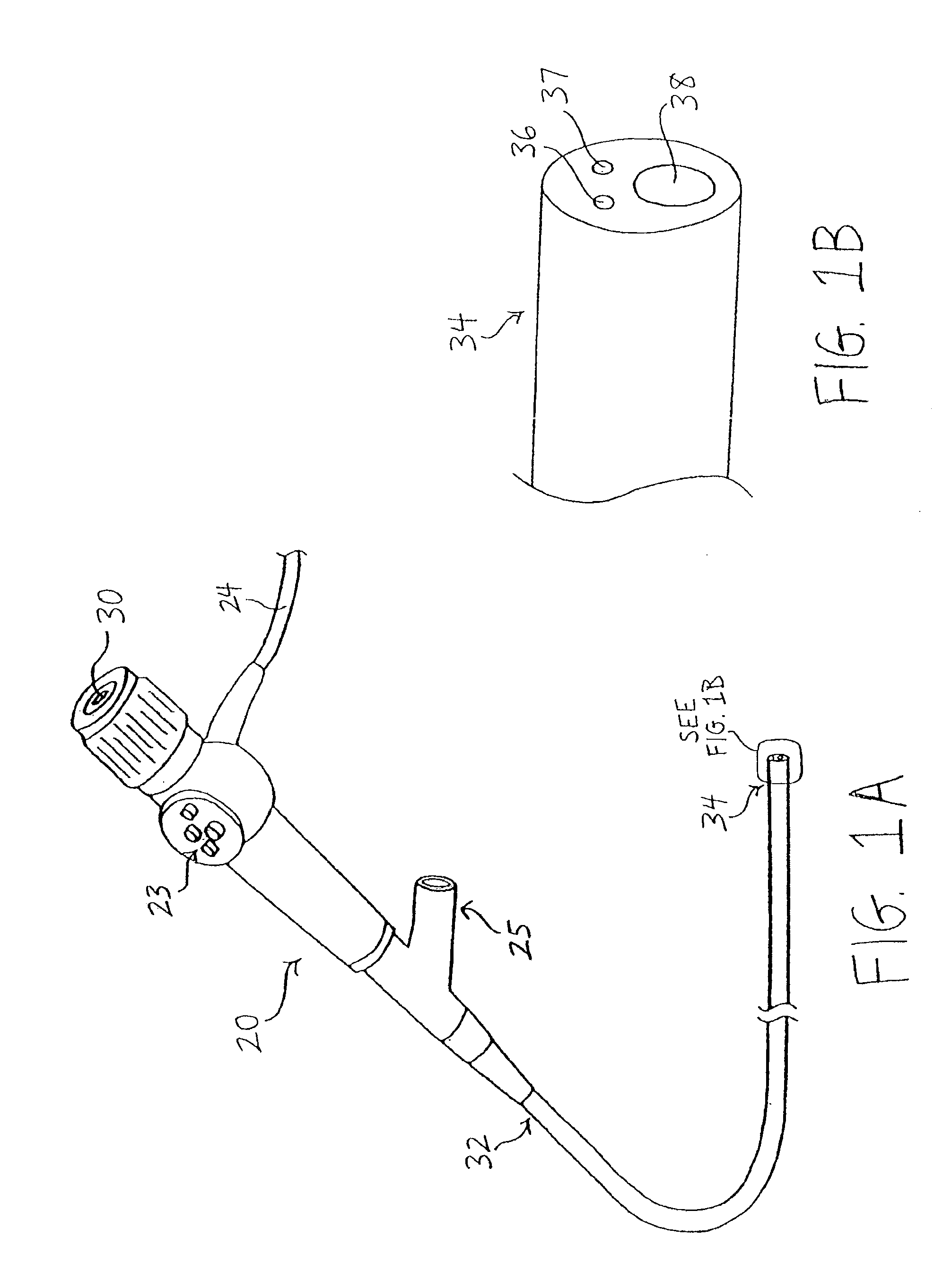
[0022]Referring now to FIGS. 1A-1B, an exemplary endoscope 20 is described, which may be used in conjunction with the tissue resection systems described below. In FIG. 1A, the exemplary endoscope 20 comprises an end-viewing endoscope of known construction and having proximal and distal regions 32 and 34, respectively. The endoscope 20 may comprise fiber optic components 36 and 37 for illuminating and capturing an image distal to the endoscope 20, as depicted in FIG. 1B. A physician may view the images distal to the endoscope 20 using an eyepiece 30. A fiber optic cable 24 may be coupled between the endoscope 20 and a suitable light source. A control section 23 may be provided to maneuver the distal region ...


PUM


Abstract

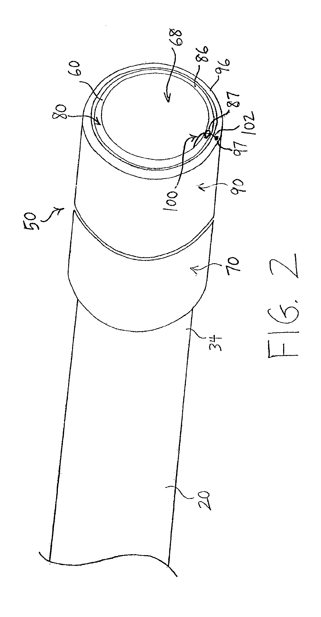
The present embodiments provide apparatus and methods for facilitating endoscopic resection of tissue. In one embodiment, the apparatus comprises a first tube having proximal and distal regions, and a working lumen disposed within at least a portion of the distal region. A second tube is disposed circumferentially around at least a portion of the distal region of the first tube. A wire has a distal end that is securely attached to a distal region of the second tube. In use, rotation of the second tube relative to the first tube causes the wire to pass circumferentially over a distal face of the first tube to incise tissue disposed within the working lumen of the first tube.

Description

PRIORITY CLAIM[0001]This invention claims the benefit of priority of U.S. Provisional Application Ser. No. 61 / 577,790, entitled “Endoscopic Systems and Methods for Resection of Tissue,” filed Dec. 20, 2011, the disclosure of which is hereby incorporated by reference in its entirety.BACKGROUND INFORMATION[0002]The present embodiments relate generally to the field of medical devices, and more particularly, to an endoscopic system for resection of tissue.[0003]There are various instances in which it may become necessary or desirable to resect a segment of a patient's tissue, for example, to obtain tissue samples for biopsies, to remove potentially harmful or cancerous localized tissue segments, and the like. In certain procedures, endoscopic mucosal resection of tissue involves using an injection needle or multi-band ligator to create a pseudo-polyp, and then subsequently resecting that tissue using an electrified snare, which is a two-part procedure.[0004]There are various other endos...

Claims


Application Information

Patent Timeline
IPC IPC(8): A61B17/32A61B18/14
CPCA61B17/32056A61B2018/144A61B2017/00269A61B2017/00296A61B2017/32004A61B2019/305A61B17/320016A61B17/32002A61B18/1477A61B18/1492A61B2018/00202A61B2018/00291A61B2018/00601A61B2018/00982A61B18/14A61B2090/035
Inventor TOOMEY, CIARANCOSTELLO, KIERAN
Owner COOK MEDICAL TECH LLC
Learn More