
Assessment of microvascular circulation

Inactive Publication Date: 2013-03-21
THE BOARD OF TRUSTEES OF THE UNIV OF ILLINOIS
View PDF3 Cites 37 Cited by
  • Summary
  • Abstract
  • Description
  • Claims
  • Application Information

AI Technical Summary

Benefits of technology

The patent describes a method for better imaging blood vessels in a target tissue by taking multiple frames of images per second. This is particularly useful when the target is moving. The method can also involve using light of a specific wavelength to enhance the imaging of blood vessels. The technical effect of this patent is improved accuracy and efficiency in blood vessel imaging.

Problems solved by technology

However, it is broadly recognized that ex vivo examination of human hemorheology, e.g., by studying blood samples, provides only limited data and typically cannot accurately characterize in vivo human circulatory conditions.
Likewise, in-situ, non-invasive measurements of human hemorheology also pose challenges in accurately describing the rheological parameters of blood flow.
For example, in vivo imaging is hindered by difficulties in tissue accessibility and mechanistic validity of the technology.

Method used


Image

  • Assessment of microvascular circulation
  • Assessment of microvascular circulation
  • Assessment of microvascular circulation

Examples


examples

[0075]Conjunctiva blood flow (BF) was determined from a sequence of image frames in the following steps: 1) image registration, 2) blood vessel centerline extraction, 3) blood vessel diameter calculation, 4) axial red blood cell velocity derivation; 5) average cross-sectional blood velocity and BF calculation. A description of each step is given below. All software and analysis algorithms were written in Matlab (The Mathworks Inc. Natick, Mass.).

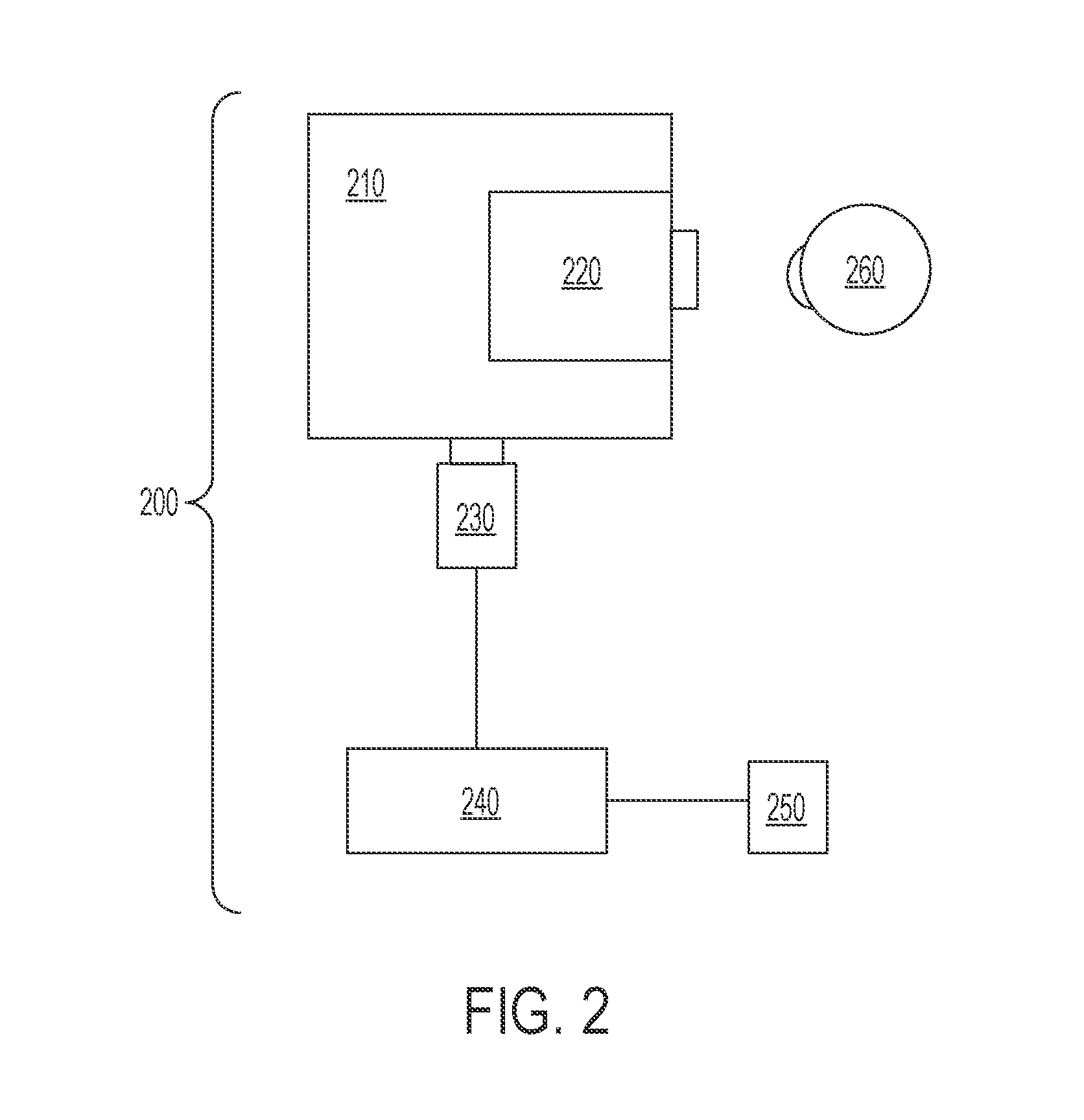
[0076]A Zeiss slit lamp biomicroscope equipped with a digital charged coupled device camera (UNIQVision Inc., Santa Clara, Calif.) was used to capture images of the human bulbar conjunctiva. A green filter with a transmission wavelength of 540±5 nm was placed in the path of the slit lamp illumination light to improve the contrast of blood vessels. The optics of the slit lamp and additional magnification optics placed in front of the camera magnified the image of conjunctiva blood vessels. The system was calibrated by capturing an image of a ...


PUM


Abstract

Methods and compositions are disclosed to quantitatively measure in vivo blood vessel diameter, blood velocity, and other flow dynamics. Such methods and compositions can optimize therapeutic interventions designed to prevent or reduce the risk of cardiovascular and blood disorders. In one aspect, the methods and apparatus involve calculating blood vessel characteristics from a two dimensional image of a blood vessel in the conjunctiva of a subject's eye. In another aspect, a series of temporal images of a blood vessel are obtained to determine blood flow properties. The apparatus can include, for example, a biomicroscope, an illuminating light source and a high speed camera to acquire the series of temporal images with the data then analyzed by a programmed processor.

Description

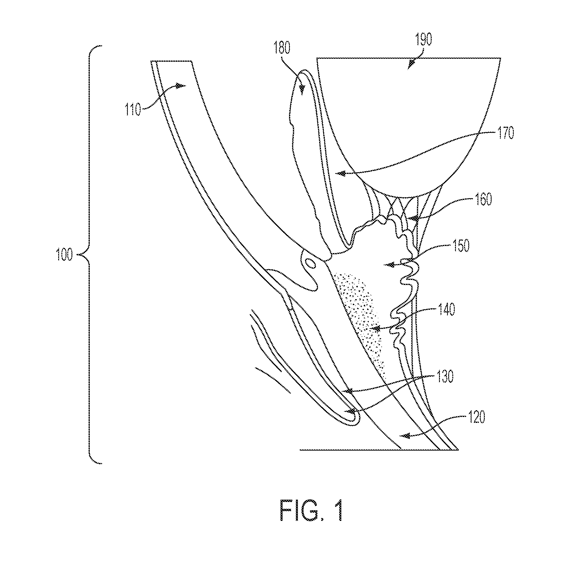
REFERENCE TO RELATED APPLICATION [0001]The present application claims priority to a provisional application entitled “Methods for Assessing Conjunctival Hemorheology of the Human Eye” filed on Nov. 30, 2009 and having Ser. No. 61 / 264,917, which is herein incorporated by reference.GOVERNMENT SPONSORSHIP[0002]This invention was made with government support under EY17918 and EY01792 awarded by the National Eye Institute. The government has certain rights in the invention.TECHNICAL FIELD[0003]The invention relates to methods and apparatus for measuring blood flow and blood flow dynamics in microvasculature.BACKGROUND OF THE INVENTION[0004]Hemorheology, the study of the flow properties of blood, has great potential for early detection or diagnosis of many illnesses, such as thromboembolisms, stroke, hyperocoagulability syndromes and blood diseases like sickle cell anemia. However, it is broadly recognized that ex vivo examination of human hemorheology, e.g., by studying blood samples, pr...

Claims


Application Information

Patent Timeline
IPC IPC(8): A61B3/14
CPCA61B3/1241A61B3/145A61B5/02007A61B3/14A61B3/10A61B5/026G01N2021/5957A61B5/113
Inventor SHAHIDI, MAHNAZWANEK, JUSTINGAYNES, BRUCE
Owner THE BOARD OF TRUSTEES OF THE UNIV OF ILLINOIS
Learn More