
Therapeutic Use of Growth Factor, and Delivery Device, Especially for the Treatment of Intimal Hyperplasia

a growth factor and intimal hyperplasia technology, applied in the field of growth factor therapy, can solve the problems of intimal hyperplasia, intimal hyperplasia, aneurysmal dilation and eventual rupture, and achieve the effect of beneficial effects on luminal size and medial and intimal thickening

Inactive Publication Date: 2012-12-06
HEALTH SCI FUNDING
View PDF3 Cites 10 Cited by
  • Summary
  • Abstract
  • Description
  • Claims
  • Application Information

AI Technical Summary

Benefits of technology

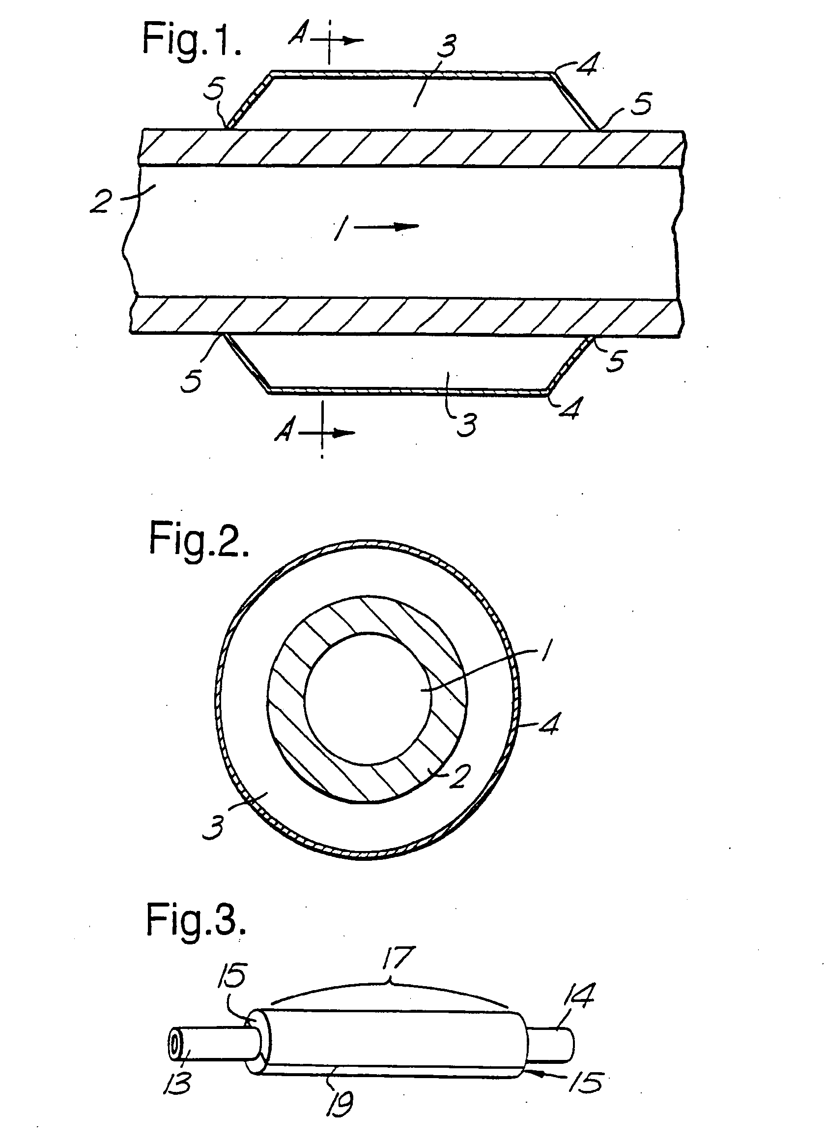
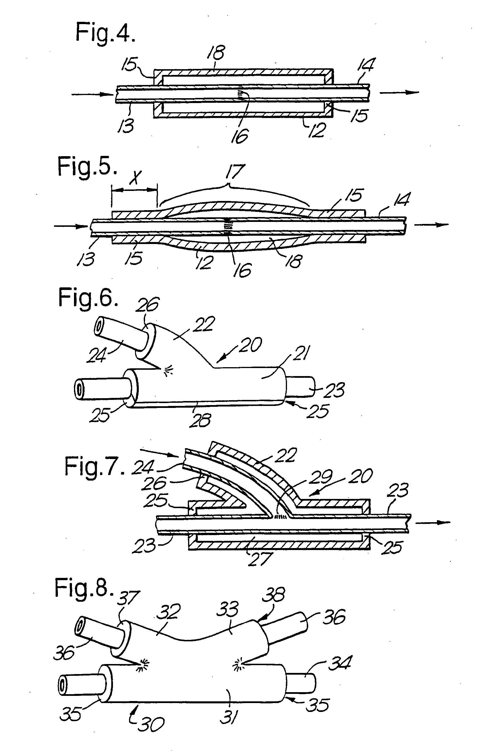
[0025]Furthermore, the new findings demonstrate that effective agents can be delivered to the exterior of the blood vessel, to treat intimal hyperplasia. This has several advantages. In particular, the therapeutic agent is not washed away from the site of the hyperplasia by blood flow as with intralumenal delivery. A delivery reservoir can be maintained around the blood vessel, and there is no need for any intralumenal manipulations which damage the endothelium of the blood vessel (and can themselves trigger intimal hyperplasia).
[0102]Some advantages of the use of implants in this way, especially collars, are: (i) they provide a delivery reservoir, allowing for sustained delivery; (ii) no intralumenal manipulations are required and the arterial endothelium remains intact; and (iii) the distortion (e.g. constriction in the case of a collar) created by the implant may enhance the efficiency of gene delivery, as explained above.

Problems solved by technology

Also, when an obstruction in a blood vessel has been cleared, intimal hyperplasia occurring after surgery may lead to the artery's becoming occluded again.
Intimal hyperplasia, whether it leads to stenosis or restenosis, remains a major problem after various surgical procedures.
Atherosclerotic cardiovascular disease is the leading cause of death in Europe and North America and prompts a highly significant morbidity consequent upon occlusion of the arterial lumen, either preventing or reducing blood flow, thrombosis superimposed upon a plaque, with possible distal embolisation, arterial wall weakening, leading to aneurysmal dilation and eventual rupture.
In the first 24 months following surgery, a very significant number of arterial bypass grafts fail (occlude).
Failure rates for ‘re-do’ procedures are even higher.
The first is recognised to occur early, within 30 days of the operation (<5%), and represents technical error (e.g. poor anastomotic technique).
However, it is those grafts that occlude between one and 24 months that form the majority of failures (<70%).
Restenosis after angioplasty can lead to even higher failure rates, from 20 to 50% in the first 6 months following the angioplasty.
Stenosis and restenosis both remain major problems after surgery.
To date, numerous methods of treating or preventing intimal hyperplasia have been tested, but none has been clinically satisfactory.
This involves the risk of infection.

Method used


Image

  • Therapeutic Use of Growth Factor, and Delivery Device, Especially for the Treatment of Intimal Hyperplasia
  • Therapeutic Use of Growth Factor, and Delivery Device, Especially for the Treatment of Intimal Hyperplasia
  • Therapeutic Use of Growth Factor, and Delivery Device, Especially for the Treatment of Intimal Hyperplasia

Examples


example 1

[0191]The effect of endothelial cell (EC)-specific VEGF gene transfer on the thickening of the intima was studied using a silicone collar inserted around carotid arteries which acted both as the agent that caused intimal smooth muscle cell growth and as a reservoir for the gene and vector. The model preserved EC integrity and permitted direct extravascular gene transfer without any intravascular manipulation.

example 1.1

Gene Transfer:

[0192]Intimal thickening was induced in the carotid arteries of thirty-two New Zealand White rabbits by inserting an inert silicone collar around the arteries under a general anaesthesia (Booth et al., Atherosclerosis (1989) 76:257-268). Gene transfer was done five days after positioning of the collar by gently opening the collar under anaesthesia and injecting 500 μl plasmid / liposome complexes into the collar (i.e. on the adventitial surface of the artery). No intravascular manipulations were involved in any steps of the studies.

Plasmid / Liposome Complexes:

[0193]Twenty-five μg pCMV5-VEGF-164 plasmid (containing mouse VEGF cDNA (Breier et al., Development (1992) 114:521-32; nucleotides 1-583) was complexed with 25 μl Lipofectin (BRL) while diluted to 500 μl with Ringer solution. Complexes were kept at room temperature at least 15 min before the gene transfer. It was determined previously that at the concentration used in the present study plasmid / Lipofectin complexes we...

example 1.2

[0198]Gene transfer was carried out as described in Example 1.1. L-NAME (70 mg / kg / d) was given to the rabbits in drinking water, starting one day before VEGF (n=5) or lacZ (n=5) gene transfer. Animals were sacrificed 7 days after gene transfer and analysed for the intima / media thickness ratio and histology as described above (Ylä-Herttuala et al., Arteriosclerosis (1986) δ: 230-236; Ylä-Herttuala et al. (1990) supra). The difference in intimal thickening was abolished.


PUM

PropertyMeasurementUnit
diameteraaaaaaaaaa
diameteraaaaaaaaaa
diameteraaaaaaaaaa

Abstract

Vascular endothelial growth factor (VEGF) has utility in the treatment of intimal hyperplasia, hypertension and atherosclerosis, and of conditions susceptible to treatment with agents that produce nitric oxide or prostacyclin. Instead of VEGF, an equivalent agent such as an agonist of VEGF receptors may be given, as may nucleic acid encoding such an agonist. The agent may successfully be administered via the adventitial surface of a blood vessel, e.g. using a device which defines a reservoir between the body wall and the vessel's adventitial surface, the reservoir being at least part-filled by a pharmaceutical formulation containing the agent to be delivered.

Description

CROSS-REFERENCE TO RELATED APPLICATION[0001]This application is a continuation-in-part of U.S. application Ser. No. 10 / 370,291, filed Feb. 19, 2003, which is a continuation of U.S. application Ser. No. 09 / 297,486, filed Jun. 14, 1999, which is the U.S. national stage of international application No. PCT / GB97 / 03015, filed Nov. 3, 1997 each of which are hereby incorporated by reference in its entirety, including all figures, nucleic acid sequences, amino acid sequences, and tables.FIELD OF THE INVENTION[0002]The present invention relates to the therapeutic use of a growth factor, and particularly to the treatment and prevention of intimal hyperplasia of blood vessels and other conditions, especially hypertension. The invention relates also to a device that can be used for delivering the active agent.BACKGROUND OF THE INVENTION[0003]Intimal hyperplasia is the increase in the number of cells between the endothelium and internal elastic lamina of a blood vessel, particularly in the intim...

Claims


Application Information

Patent Timeline
Patent Type & Authority Applications(United States)
IPC IPC(8): A61K35/12A61P7/00A61M5/00
CPCA61F2250/0067A61K48/00A61K48/005C12N15/86A61K38/1866C12N2710/10371C12N2740/13043C12N2740/13045C12N2710/10343A61P7/00A61P9/00A61P9/12
Inventor MARTIN, JOHN FRANCISYLA-HERTTUALA, SEPPOBARKER, STEPHEN GEORGE EDWARD
Owner HEALTH SCI FUNDING
Learn More