Method and System for Regression-Based 4D Mitral Valve Segmentation From 2D+t Magnetic Resonance Imaging Slices

a regression-based, mitral valve technology, applied in image analysis, medical science, image enhancement, etc., can solve the problems of no established acquisition protocol for extracting 4d anatomical and function information regarding the heart valve, less research on extracting the heart valve from cmr data, and limiting the capabilities of accurate 4d anatomical and functional analysis of the hear

Active Publication Date: 2012-09-13
SIEMENS HEALTHCARE GMBH
View PDF4 Cites 6 Cited by
  • Summary
  • Abstract
  • Description
  • Claims
  • Application Information

AI Technical Summary

Problems solved by technology

However, the 2D slice based acquisition of CMR limits the capabilities for accurate 4D anatomical and functional analysis of the heart due to long throughput times and the need for protracted study.
Although many groups have studied 4D heart chamber acquisition and segmentation, with a focus on the left ventricle, to overcome the 2D acquisition limitations of CMR, there is stil

Method used


Image

  • Method and System for Regression-Based 4D Mitral Valve Segmentation From 2D+t Magnetic Resonance Imaging Slices
  • Method and System for Regression-Based 4D Mitral Valve Segmentation From 2D+t Magnetic Resonance Imaging Slices
  • Method and System for Regression-Based 4D Mitral Valve Segmentation From 2D+t Magnetic Resonance Imaging Slices

Examples


Embodiment Construction

[0014]The present invention relates to regression-based segmentation of the mitral valve (MV) from 2D+t magnetic resonance (MR) images. Embodiments of the present invention are described herein to give a visual understanding of the mitral valve segmentation method. A digital image is often composed of digital representations of one or more objects (or shapes). The digital representation of an object is often described herein in terms of identifying and manipulating the objects. Such manipulations are virtual manipulations accomplished in the memory or other circuitry / hardware of a computer system. Accordingly, it is to be understood that embodiments of the present invention may be performed within a computer system using data stored within the computer system.

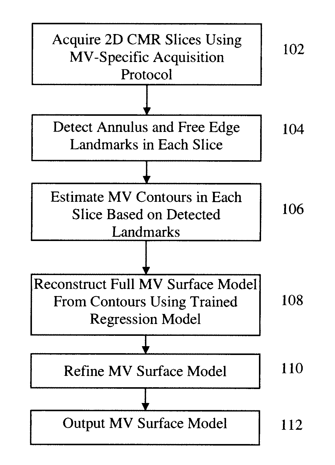
[0015]In order to accurately reconstruct the surface of the MV from incomplete data, embodiments of the present invention introduce a regression-based method for segmenting a complete MV surface model. In particular, a set of 3...


PUM


Abstract

A system and method for regression-based segmentation of the mitral valve in 2D+t cardiac magnetic resonance (CMR) slices is disclosed. The 2D+t CMR slices are acquired according to a mitral valve-specific acquisition protocol introduced herein. A set of mitral valve landmarks is detected in each 2D CMR slice and mitral valve contours are estimated in each 2D CMR slice based on the detected landmarks. A full mitral valve model is reconstructed from the mitral valve contours estimated in the 2D CMR slices using a trained regression model. Each 2D CMR slice may be a cine image acquired over a full cardiac cycle. In this case, the segmentation method reconstructs a patient-specific 4D dynamic mitral valve model from the 2D+t CMR image data.

Description

[0001]This application claims the benefit of U.S. Provisional Application No. 61 / 451,025, filed Mar. 9, 2011, the disclosure of which is herein incorporated by reference.BACKGROUND OF THE INVENTION[0002]The present invention relates to cardiac magnetic resonance (MR) imaging, and more particularly, to 4D mitral valve segmentation from 2D+t MR slices.[0003]Cardiac MR imaging (CMR) has recently emerged as the new gold standard for characterizing cardiac masses and evaluating cardiac function and anatomy. The multi-plan ability of CMR to acquire tomographic images in any plane, the capability to measure blood flow velocity in all three dimensions within a single slice, and the use of non-ionizing radiation are significant advantages of CMR over other imaging modalities. Clinical studies have proven that CMR is well suited for the complex evaluation of the mitral valve by comparing mitral valve measurements extracted from CMR data with those extracted from computed tomography (CT) data ...

Claims


Application Information

Patent Timeline
IPC IPC(8): A61B5/055
CPCG06T7/0083G06T2207/10088G06T2207/20081G06T2207/20076G06T2207/30048G06T7/12
Inventor IONASEC, RAZVAN IOANVITANOVSKI, DIMETSYMBAL, ALEXEYFUNKA-LEA, GARETHCOMANICIU, DORINGREISER, ANDREASMUELLER, EDGAR
Owner SIEMENS HEALTHCARE GMBH
Why Eureka
Try Eureka