Spectral doppler ultrasound imaging device and method for controlling same

a technology imaging device, which is applied in tomography, applications, instruments, etc., can solve the problems of affecting the accuracy and affecting the user's workflow. , to achieve the effect of simplifying the use of spectral doppler ultrasound imaging devi

Inactive Publication Date: 2012-03-08
KONINKLIJKE PHILIPS ELECTRONICS NV
View PDF1 Cites 6 Cited by
  • Summary
  • Abstract
  • Description
  • Claims
  • Application Information

AI Technical Summary

Benefits of technology

[0011]It is an object of the invention to provide a method for controlling a spectral Doppler ultrasound imaging device adapted for operating both in a spectrum-live Doppler measurement mode and in an image-live measurement mode wherein the method may simplify the use of the spectral Doppler ultrasound imaging device. It is another object of the invention to provide a spectral Doppler ultrasound imaging device adapted for operating in accordance to such simplifying method, a computer program product adapted to control, when executed on a computer, a spectral Doppler ultrasound imaging device in accordance with such simplifying method and a computer-readable medium having such computer program product stored thereon.
[0018]Embodiments of the invention deal with spectral Doppler ultrasound imaging. Such spectral Doppler ultrasound imaging may allow accurate quantitative measurement of e.g. blood flow velocities, compared to other kinds of imaging modalities.
[0023]In other words, the proposed ultrasound imaging system may automatically switch to the image-live measurement mode preferably exactly at those instances when a user may need the color or grey-scale 2D image-live, namely then when the user wants to change a sampled region corresponding to a region of interest. Accordingly, the user may actively indicate a desired change of the sampled region for example by indicating such change on a trackball or angle knob or by moving the ultrasound transducer of the imaging device and the imaging device will detect such indicated change and automatically switch to the image-live measurement mode such that the user may see and, if necessary, correct the position or angle of the sampled region within the 2D ultrasound image. Thereby, tedious efforts for repeatedly toggling between image-live and spectrum-live measurement modes may be prevented simplifying the use of the ultrasound imaging device. Furthermore, the imaging device may be in the spectrum-live measurement mode most of the time allowing high quality spectrum analysis and is only interrupted by switching into the image-live measurement mode for the case, that a change of a sampled region is detected.
[0024]According to an embodiment of the present invention, the method further comprises automatically switching to the spectrum-live Doppler measurement mode a predetermined duration after the detection of the change of the sampled region. For example, such predetermined duration may be one second. In other words, upon detection of a change of the sampled region, the device automatically switches from the spectrum-live measurement mode to the image-live measurement mode and then, after e.g. one second, back to the spectrum-live measurement mode. Accordingly, the imaging device is within the spectrum-live measurement mode most of the time and is only interrupted for a short period of time by the image-live measurement mode. This may improve the quality of the spectrum-live measurements.
[0026]According to a further embodiment of the present invention, the Doppler ultrasound imaging device is controlled to operate at least in one of the spectrum-live Doppler measurement mode and the image-live measurement mode in a full frame rate mode. Preferably, the device operates in the full frame rate mode in both, spectrum-live and image-live measurement modes. Therein, the term “full frame rate mode” may indicate that images or spectra are acquired continuously and not only as a single frame update for each change of a sampled region. Furthermore, the imaging device preferably operates in a real-time mode in both, the spectrum-live and the image-live measurement modes. Therein, the term “real-time” may indicate that there is only a negligible delay in time between a real motion in the sampled region and its acquisition and display by the imaging device. Accordingly, in both measurement modes, the device may operate with the highest possible imaging quality and compromises are avoided.

Problems solved by technology

The toggling between the spectrum-live mode and the image-live mode may complicate a user's workflow during the Doppler part of an ultrasound exam due to the conventionally necessary repetitive use of the ENTER / UPDATE key to manually toggle between the spectrum-live and image-live mode for adjusting the position or angle of the sampled region.
However, this does not show the flow dynamics, is different or unlikely to catch a jet or systolic pulse, may interrupt the several seconds needed for Doppler spectrum measurements and may waste most of an update time with slewing and settling a transmit voltage.
However, these techniques inevitably involve compromises in velocity scale, frame rate, sensitivity, artefacts and / or measurement accuracy.
However, since such techniques depend on acquiring image-live measurements they have similar issues of switching between spectrum-live and image-live mode as described further above and may compromise both.

Method used


Image

  • Spectral doppler ultrasound imaging device and method for controlling same
  • Spectral doppler ultrasound imaging device and method for controlling same
  • Spectral doppler ultrasound imaging device and method for controlling same

Examples


Embodiment Construction

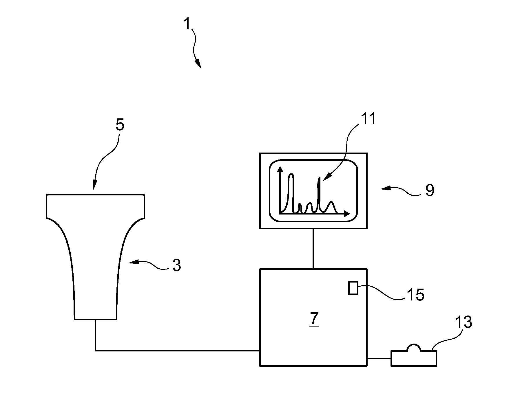
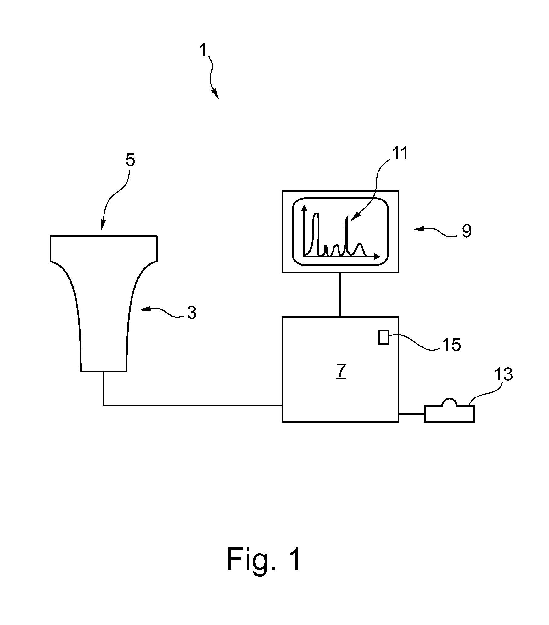
[0034]In FIG. 1, a spectral Doppler ultrasound imaging device 1 according to an embodiment of the present invention is schematically depicted. An ultrasound transducer 3 comprises an ultrasound transceiver face 5 from which ultrasound waves can be emitted into a patient's body and which may then detect reflected echoes. The transducer 3 is connected to a control device 7. The control device 7 may receive the detected echo signals from the transducer 3 and, based thereon, may display corresponding ultrasound images on a display 9. In a first operation mode, the control device 7 may control the transducer 3 to operate in an image-live measurement mode to provide color or grey-scale two-dimensional (2D) or three-dimensional (3D) ultrasound images to be displayed on the display 9. In a second mode, the control device 7 may control the transducer 3 to operate in a spectrum-live Doppler measurement mode and e.g. a quantitative analysis of velocities of motions within a sampled region may ...


PUM


Abstract

A method for controlling a spectral Doppler ultrasound imaging device adapted for operating both in a spectrum-live Doppler measurement mode (S3) and in an image-live measurement mode (S1, S6) is proposed. In accordance with the present invention, the method comprises automatically switching to the image-live measurement mode (S6) upon detection (S5) of a change of a sampled region. A change of a sampled region may be indicated by an operator by actuating a sampled region control device such as a trackball, angle knob or sample gate controller and, upon such actuation, the spectral Doppler ultrasound imaging device may automatically switch to the image-live measurement mode (S6) to allow for adapting or correcting of a position and / or orientation of the sampled region. After the sampled region has been positionally stable for a predetermined duration, the spectral Doppler ultrasound imaging device may be switched back (S7) to the spectrum-live measurement mode (S3). Thereby, unnecessary manual toggling between different measurement modes may be avoided and quantitative spectrum-live Doppler measurement (S3) is enabled being interrupted only when necessary, i.e. when a sampled region is changed.

Description

FIELD OF THE INVENTION[0001]The invention relates to a method for controlling a spectral Doppler ultrasound imaging device adapted for operating both in a spectrum-live Doppler measurement mode and in an image-live measurement mode. Furthermore, the invention relates to such spectral Doppler ultrasound imaging device being adapted for performing the inventive method, to a computer program product for accordingly controlling a spectral Doppler ultrasound imaging device when executed on a computer and a computer-readable medium having such computer program product stored thereon.BACKGROUND OF THE INVENTION[0002]Ultrasound imaging devices have been developed for qualitatively and quantitatively imaging both static structures within a sampled region and dynamic motions within the sampled region. Therein, ultrasound is emitted towards the sampled region and echoes reflected within the sampled region are detected. The sampled region, sometimes also referred to as region of interest, may b...

Claims


Application Information

Patent Timeline
IPC IPC(8): A61B8/00
CPCA61B8/06A61B8/13A61B8/488G01S15/8979G01S7/52066G01S7/52084G01S7/52063
Inventor CLARK, DAVID
Owner KONINKLIJKE PHILIPS ELECTRONICS NV
Why Eureka
Try Eureka