
Devices for creating passages and sensing for blood vessels

a technology of blood vessels and devices, applied in the field of devices for creating passages and sensing for blood vessels, can solve the problems of copd-afflicted individuals also facing muscle strength loss, little hope of recovery, and inability to perform common daily activities. achieve the effect of greater pressur

Inactive Publication Date: 2011-12-15
BRONCUS MEDICAL
View PDF1 Cites 43 Cited by
  • Summary
  • Abstract
  • Description
  • Claims
  • Application Information

AI Technical Summary

Benefits of technology

The invention is a device and method for creating passages and removing tissue while sensing blood vessels in the body, particularly in the lungs. The device includes an elongate member with a tissue piercing member and an ultrasound transducer assembly that can move relative to each other. The device can create passages and remove tissue while sensing blood vessels in the area. The device can also include a blunt tip and an expandable member, such as a balloon, for dilating tissue. The device can communicate with an analyzing device or control unit to determine the presence or absence of blood vessels based on the reflected signals. The invention can be used in the lungs to treat lung tissue by creating openings and sensing blood vessels.

Problems solved by technology

Those inflicted with COPD face disabilities due to the limited pulmonary functions.
Usually, individuals afflicted by COPD also face loss in muscle strength and an inability to perform common daily activities.
Since the damage to the lungs is irreversible, there is little hope of recovery.
Most times, the physician cannot reverse the effects of the disease but can only offer treatment and advice to halt the progression of the disease.
However, lung reduction surgery is an extremely traumatic procedure which involves opening the chest and thoracic cavity to remove a portion of the lung.
If the entire lung is emphysematous, however, removal of a portion of the lung removes gas exchanging alveolar surfaces, reducing the overall efficiency of the lung.
Lung volume reduction surgery is thus not a practical solution for treatment of emphysema where the entire lung is diseased.
Both bronchodilator drugs and lung reduction surgery fail to capitalize on the increased collateral ventilation taking place in the diseased lung.

Method used


Image

  • Devices for creating passages and sensing for blood vessels
  • Devices for creating passages and sensing for blood vessels
  • Devices for creating passages and sensing for blood vessels

Examples


Embodiment Construction

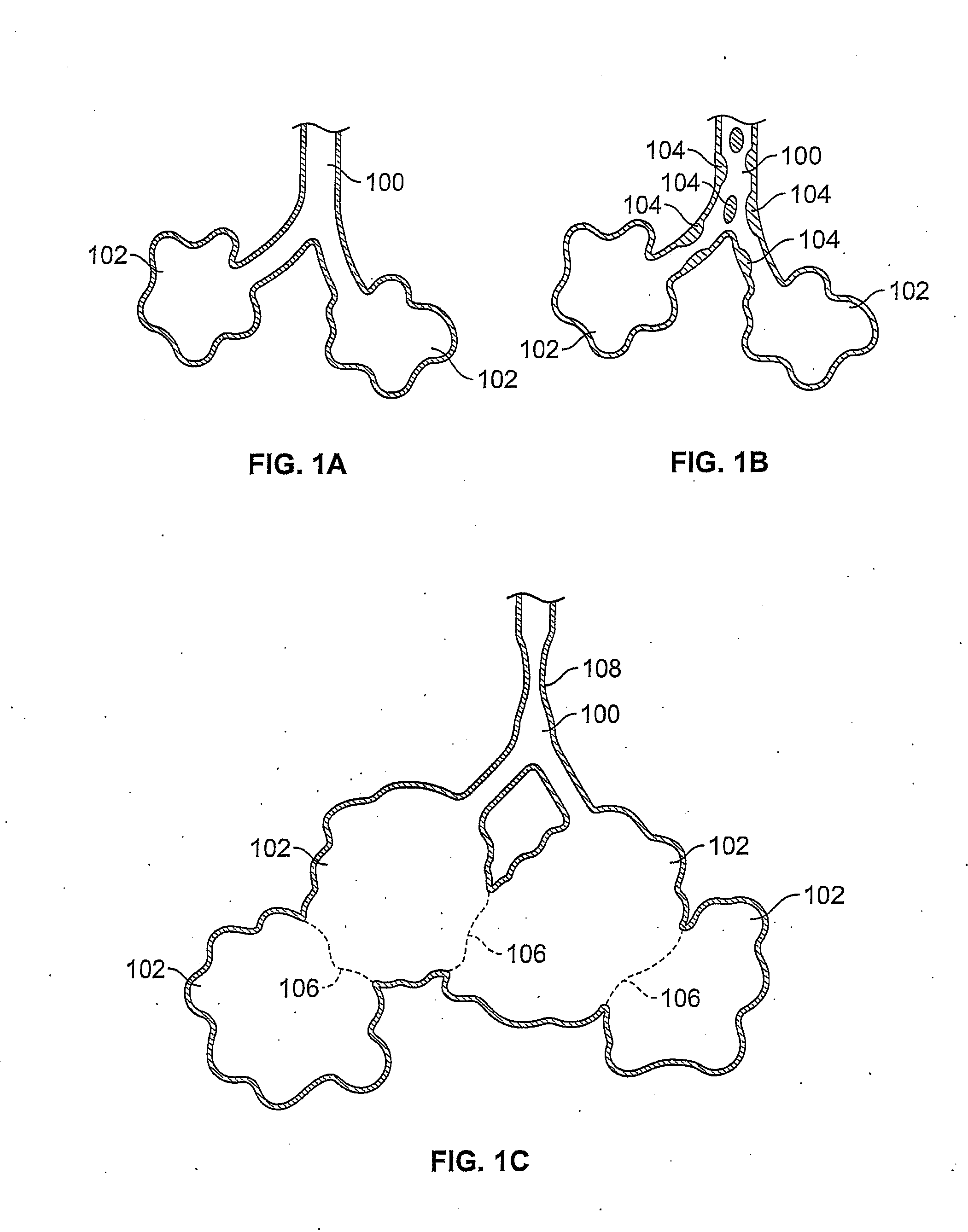
[0031]FIG. 1A shows a simplified illustration of a natural airway 100 which eventually branches to a blood gas interface 102. FIG. 1B illustrates an airway 100 and blood gas interface 102 in an individual having COPD. The obstructions 104 (e.g., excessive mucus resulting from COPD, see above) impair the passage of gas between the airways 100 and the interface 102. FIG. 1C illustrates a portion of an emphysematous lung where the blood gas interface 102 expands due to the loss of the interface walls 106 which have deteriorated due to a bio-chemical breakdown of the walls 106. Also depicted is a constriction 108 of the airway 100. It is generally understood that there is usually a combination of the phenomena depicted in FIGS. 1A-1C. More usually, the states of the lung depicted in FIGS. 1B and 1C are often found in the same lung.

[0032] As will be explained in greater detail below, the production and maintenance of collateral openings or channels through airway walls permits expired a...


PUM


Abstract

Devices and methods are disclosed for creating passages in tissue and detecting blood vessels in and around the passages. The devices may be used to create channels for altering gaseous flow within a lung to improve the expiration cycle of an individual, particularly individuals having Chronic Obstructive Pulmonary Disease (COPD). In addition, the devices may be used to sample tissue during biopsy or other medical procedures where perforating a blood vessel could result in injury to a patient.

Description

FIELD OF THE INVENTION [0001] The invention is directed to devices for creating passages in tissue and detecting blood vessels in and around the passages. The device may be used to create channels for altering gaseous flow within a lung to improve the expiration cycle of an individual, particularly individuals having Chronic Obstructive Pulmonary Disease (COPD). In addition, the device may be used to sample tissue during biopsy or other medical procedures where perforating a blood vessel could result in injury to a patient. BACKGROUND OF THE INVENTION [0002] The American Lung Association (ALA) estimates that nearly 16 million Americans suffer from chronic obstructive pulmonary disease (COPD) which includes diseases such as chronic bronchitis, emphysema, and some types of asthma. The ALA estimated that COPD was the fourth-ranking cause of death in the U.S. The ALA estimates that about 14 million and 2 million Americans suffer from emphysema and chronic bronchitis respectively. [0003]...

Claims


Application Information

Patent Timeline
Patent Type & Authority Applications(United States)
IPC IPC(8): A61B17/34
CPCA61B17/32053A61B17/32093A61B2017/3413A61B2017/00778A61B2017/320048A61B2017/00252
Inventor ROSCHAK, EDMUND J.KEAST, THOMAS M.MASSETTI, PATRICK JOHN
Owner BRONCUS MEDICAL
Learn More