Nucleic acid isolation in preserved whole blood
a technology of whole blood and nucleic acid, which is applied in the field of dna isolation from biological samples, can solve the problems of dna shearing, failure to remove unwanted materials from dna samples, and difficulty if not impossible to use samples as diagnostic tools
- Summary
- Abstract
- Description
- Claims
- Application Information
AI Technical Summary
Benefits of technology
Problems solved by technology
Method used
Image
Examples
example 1
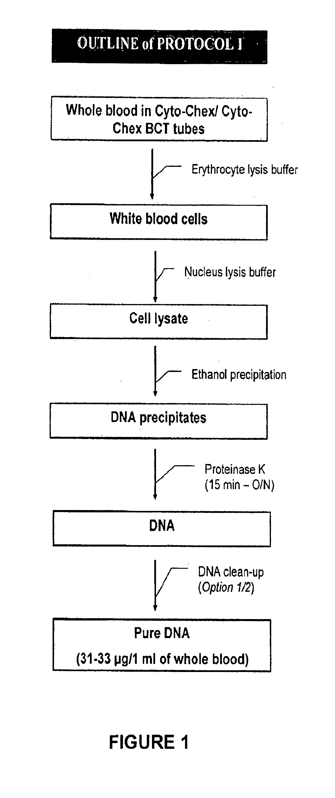
[0033]Mix 1 ml of whole blood with 5 ml of erythrocyte lysis buffer in a 15 ml centrifuge tube. Vortex briefly and incubate for 10 to 20 minutes on ice to lyse the red blood cells. Centrifuge at 1000 rpm for 10 minutes at 4-9° C. and discard the supernatant. Add 2 ml of erythrocyte lysis buffer to cell pellet. Re-suspend the cells by vortexing briefly at high speed. Centrifuge at 1000 rpm for 10 minutes at 4-9° C. and discard supernatant. It is important to remove the supernatant as much as possible to avoid incomplete lysis of white blood cells. If desired, the process can be stopped at this point and the cell pellet can be maintained at −80° C. for many months. To proceed, prepare a white blood cell lysis buffer by adding β-mercaptoethanol to nucleus lysis buffer in a 1:100 dilution ratio. Mix well by inverting. Add the white blood cell lysis buffer to the cell pellet and vortex until no cell clumps are visible. Transfer the cell lysate to a clean microcentrifuge tube. Add 1 / 10 vo...
example 2
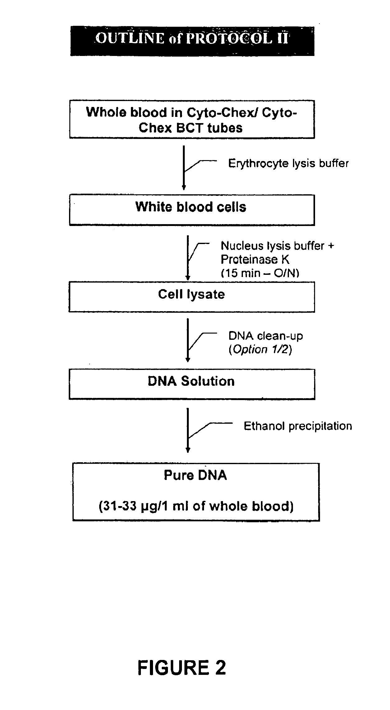
[0034]Repeat all steps of Example 1. Continue by adding an equal volume of phenol:chloroform:isoamyl alcohol (25:24:1 saturated with 10 mM Tris, pH 8.0 or 1 mM EDTA) to DNA solution. Vortex for 10 seconds and centrifuge at 12,000 rpm at room temperature for 5 minutes. Take the aqueous phase containing the DNA and transfer to a new tube. Add 1 / 10 volume ( 1 / 10 of cell lysate) of 5M NaCl to the cell lysate and mix well by inverting. Add 1 volume (equal volume of cell lysate) of 100% isopropanol to the cell lysate and mix well by inverting. Incubate at −20° C. for a minimum of 30 minutes. Again, the process can be stopped at this point as the DNA is considered stable. To proceed, centrifuge at 4° C. and 13,000 rpm for 20-30 minutes. Pour off the supernatant and discard. Add 1 ml 70% ethanol to the pellet, vortex for 10 seconds and centrifuge at 4° C. and 13,000 rpm for 10 minutes. Pour off the supernatant and discard. Repeat the addition of ethanol and centrifuging. Drain the microcent...
example 3
[0035]Repeat all steps of Example 1. Add 67 μl of protein precipitation solution to DNA solution. Vortex to mix and incubate on ice for 5 minutes. Centrifuge at 13,000 rpm at room temperature for 10 minutes. Remove the supernatant to a clean microcentrifuge tube. Add 1 / 10 volume ( 1 / 10 of cell lysate) of 5M NaCl to the cell lysate and mix well by inverting. Add 1 volume (equal volume of cell lysate) of 100% isopropanol to the cell lysate and mix well by inverting. Incubate at −20° C. for a minimum of 30 minutes. Again, the process can be stopped at this point as the DNA is considered stable. To proceed, centrifuge at 4° C. and 13,000 rpm for 20-30 minutes. Pour off the supernatant and discard. Add 1 ml 70% ethanol to the pellet, vortex for 10 seconds and centrifuge at 4° C. and 13,000 rpm for 10 minutes. Pour off the supernatant and discard. Repeat the addition of ethanol and centrifuging. Drain the microcentrifuge tube and allow DNA pellet to air dry in the open tube. Add 200 μl TE...
PUM
| Property | Measurement | Unit |
|---|---|---|
| concentration | aaaaa | aaaaa |
| temperature | aaaaa | aaaaa |
| temperature | aaaaa | aaaaa |
Abstract
Description
Claims
Application Information
 - R&D
- Intellectual Property
- Life Sciences
- Materials
- Tech Scout
- Unparalleled Data Quality
- Higher Quality Content
- 60% Fewer Hallucinations
Browse by: Latest US Patents, China's latest patents, Technical Efficacy Thesaurus, Application Domain, Technology Topic, Popular Technical Reports.
© 2025 PatSnap. All rights reserved.Legal|Privacy policy|Modern Slavery Act Transparency Statement|Sitemap|About US| Contact US: help@patsnap.com


