Microtrench and tumour proliferation assay

Inactive Publication Date: 2011-07-14
UNIV COLLEGE CARDIFF CONSULTANTS LTD
View PDF1 Cites 16 Cited by
  • Summary
  • Abstract
  • Description
  • Claims
  • Application Information

AI Technical Summary

Benefits of technology

[0034]A cell exposed to a test agent may be observed after a given time, e.g., after one hour, five hours, 10 hours, 24 hours, 1 week. The time interval selected would preferably be in excess of a cell cycle period (nominally >22 h for U-2 OS cells) to observe cell cycle perturbations, greater than two cell cycles (nominally >44 h for U-2 OS cells) to observe minimal clonogenic potential, over shorter time frames to assess the induction of acute cell death, or over very short intervals (e.g., <5 h) to assess cell movement. The skilled person would readily be able to determined appropriate time intervals according to the cell type used.
[0147]Detection of nanoparticle (conveniently describing a particle having one or more dimensions of the order of 100 nm or less) toxicity with the advantage that cells carrying a toxic load of particles and undergoing proliferation arrest can be located for definitive analysis using electron microscopy or other high resolution imaging approaches.

Problems solved by technology

A resultant problem is that the cell is in a 3-D cluster in these circumstances, making individual cell analysis difficult, fixation problematic and divisional history effectively impossible to determine.
A cell population can contain a range of proliferation components (i.e., not all cells behave the same) causing problems for identifying and determining the proportion of cells that have not undergone cell division as distinct from those that have undergone rounds of division to produce cell lineages, particularly if the division event has not been visually ‘captured’ during the analysis.
Further, inherent or induced differences in proliferation rates between cells can impose asynchrony, demanding kinetic analyses of single cells.
Motility can cause a problem for cell re-identification when re-visiting the original location of a cell at a later time point and furthermore potential removal of a cell from the enumerated fraction (see also cell incursion).
Cell loss—this causes a problem for enumeration of a fraction of cells that may be lost (e.g., as the result of cell death) in mixed populations which are also undergoing cell proliferation.
Cell incursion—relating to the adventitious appearance of cells from outside a field undergoing analysis / observation (e.g., via cell motility or a process of detachment and reattachment) effectively resulting in a ‘contamination’ of a field of interest increasing the noise in the system.
This can cause problems for cell identification particularly when applying image analysis or segmentation algorithms, whereby cell shape changes may act to confound analysis procedures or reduce their efficiency.
Microniche exhaustion of growth and survival factors—when cells are constrained within diffusion-limited circumstances (e.g., in a micropocket), there is the potential to limit access to vital molecules in the supporting medium and access to analytical reagents.
Further, separation of cells into separated micro-niche areas effectively reduces local cell density and may thereby reduce the ability of cell cultures to ‘condition’ to their overall environment.
This approach leads to further complications with respect to increased cell interactions that can compromise assay performance.
Another common solution is to increase the number of observed views of a population although this leads to an increase in the amount of data collected, which must then be stored.
Uncertainty in terms of progenitor identification and descendant relationships introduces noise into an analytical system.
Such microwells could be used for adherent cells, depending upon the biocompatibility of the substrate but do not intrinsically control cell contacts.
A resultant problem is that the cell now undergoes changes associated with the adhesion responses and such selective capture does not reveal divisional history.
However, within such micro-well formats, cell orientation is not ordered to enhance the means of analysis (temporal order of events) or to allow the reporting of a cell with a deduced divisional history.
Clonogenic potential may be limited according to cell type.
According to the type of culture, cells can become limited in their proliferation potential by local conditions such as growth factor exhaustion and the operation of contact inhibition.
This approach has the draw-back that a clonogenic assay typically requires 1-3 weeks of incubation to stabilise a macroscopic colony and uses low plating densities that can both compromise clonogenicity and restrict the ability to identify progenitors in sparse fields.
Problems also relate to the exhaustion of medium during cultivation and the production of satellite colonies due to cell detachment and re-attachment at a different location.
Some highly motile cells can act to diffuse colony structure and make colony recognition problematic.
The approach is labour-intensive for manual and semi-manual tracking and can potentially result in deleterious effects on cell cultures due to the high frequency of light exposures required to capture and therefore register a transient event such as cell division.
Time-lapse imaging typically generates large amounts of image-based information which results in problems for short- and long-term data storage, analysis and access.
Furthermore, the acquisition platform is often continuously occupied, resulting in limitations for assay throughput.

Method used


Image

  • Microtrench and tumour proliferation assay
  • Microtrench and tumour proliferation assay
  • Microtrench and tumour proliferation assay

Examples


Embodiment Construction

Selection of Substrate and Topological Conditions to Influence Cell Adhesion

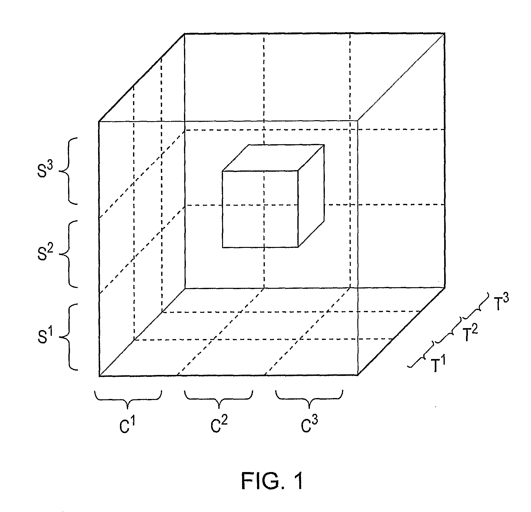
[0174]FIG. 1 shows conceptually, by way of example, how three variable influences (degrees of freedom) may be combined to describe conditions suitable for a cell-based assay on a microfeatured surface. The quality S represents substrate characteristics that can be varied according to different degrees S1, S2, S2 or Sn. The quality C represents cell-associated characteristics that can be varied or are presented according to different degrees C1, C2, C2 or Cn. The quality T represents topological characteristics of the substrate that can be varied according to different degrees T1, T2, T2 or Tn. By way of example, S′ could represent tissue culture (TC) plastic, S2 an unmodified PDMS substrate and S3 a modified PDMS substrate. By way of example, C1-n could represent a cellular characteristic such as the variable expression of a cell surface adhesion molecule. By way of example, T1-n could represent the variable...


PUM


Abstract

There is provided a cell culture microtrench being defined on or in a surface of a substrate, wherein the ratio of the width of the microtrench to the maximum length of the short axis of a cell type of interest is about 6 or preferably less, the length of the short axis of the cell type being measured when a cell is in detached or suspended form. There is also provided an array comprising such a microtrench and uses of such microtrenches, including cell-based assays.

Description

FIELD OF THE INVENTION[0001]The invention relates to the use of a cell culture microtrench having dimensions which allow attachment of cells to the surface of the trench and constrain the location and behaviour of cells for analytical advantage. The invention also relates to such a microtrench, arrays of such trenches with varying dimensions to provide for selection of optimal assay characteristics for a user-defined purpose, and arrays of trenches with the same dimensions to facilitate the scaling up and efficiency of a given assay. The trenches and / or arrays can be readily incorporated into convenient methods of cellular analysis in research, diagnostics and screening assays.BACKGROUNDConfounding Factors in Cell-Based Assays[0002]The determination of the abilities of cultured cells to divide, grow, survive and display molecular features or behavioural characteristics underpin many biological analyses in basic cellular research, the evaluation of cell therapeutic preparations, biot...

Claims


Application Information

Patent Timeline
IPC IPC(8): G01N33/50C12Q1/02C12M3/00C40B40/02C12N5/09
CPCG01N33/5011G01N33/5005
Inventor SMITH, PAUL J.ERRINGTON, RACHEL J.SUMMERS, HUWCHAPPELL, SALLY
Owner UNIV COLLEGE CARDIFF CONSULTANTS LTD
Why Eureka
Try Eureka