
Pluripotent stem cell that can be isolated from body tissue

a stem cell and body tissue technology, applied in the field of body tissuederived pluripotent stem cells, can solve the problems of unreliable stem cell differentiation, limited pluripotency, and low tissue regenerative capacity, and achieve the effect of reducing the number of pluripotent stem cells in mature mammalian bodies and reducing the number of pluripotent stem cells

Active Publication Date: 2011-03-24
DEZAWA MARI +3
View PDF6 Cites 64 Cited by
  • Summary
  • Abstract
  • Description
  • Claims
  • Application Information

AI Technical Summary

Benefits of technology

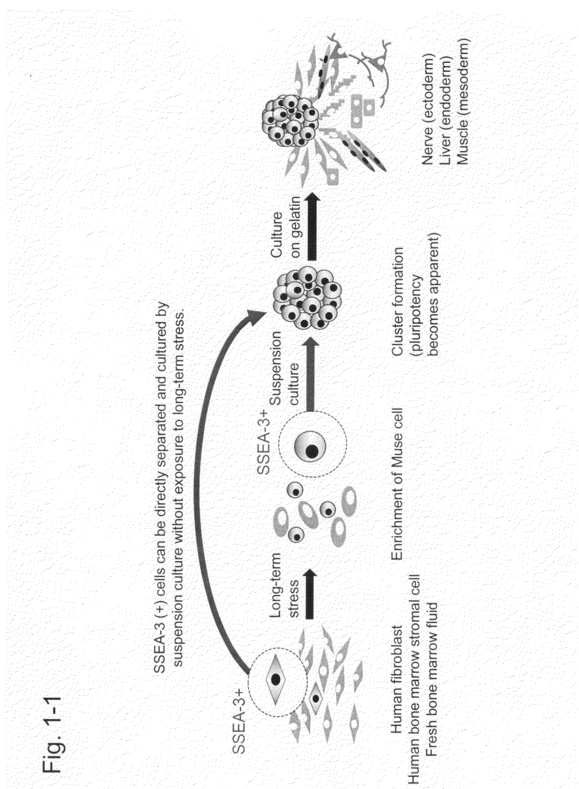
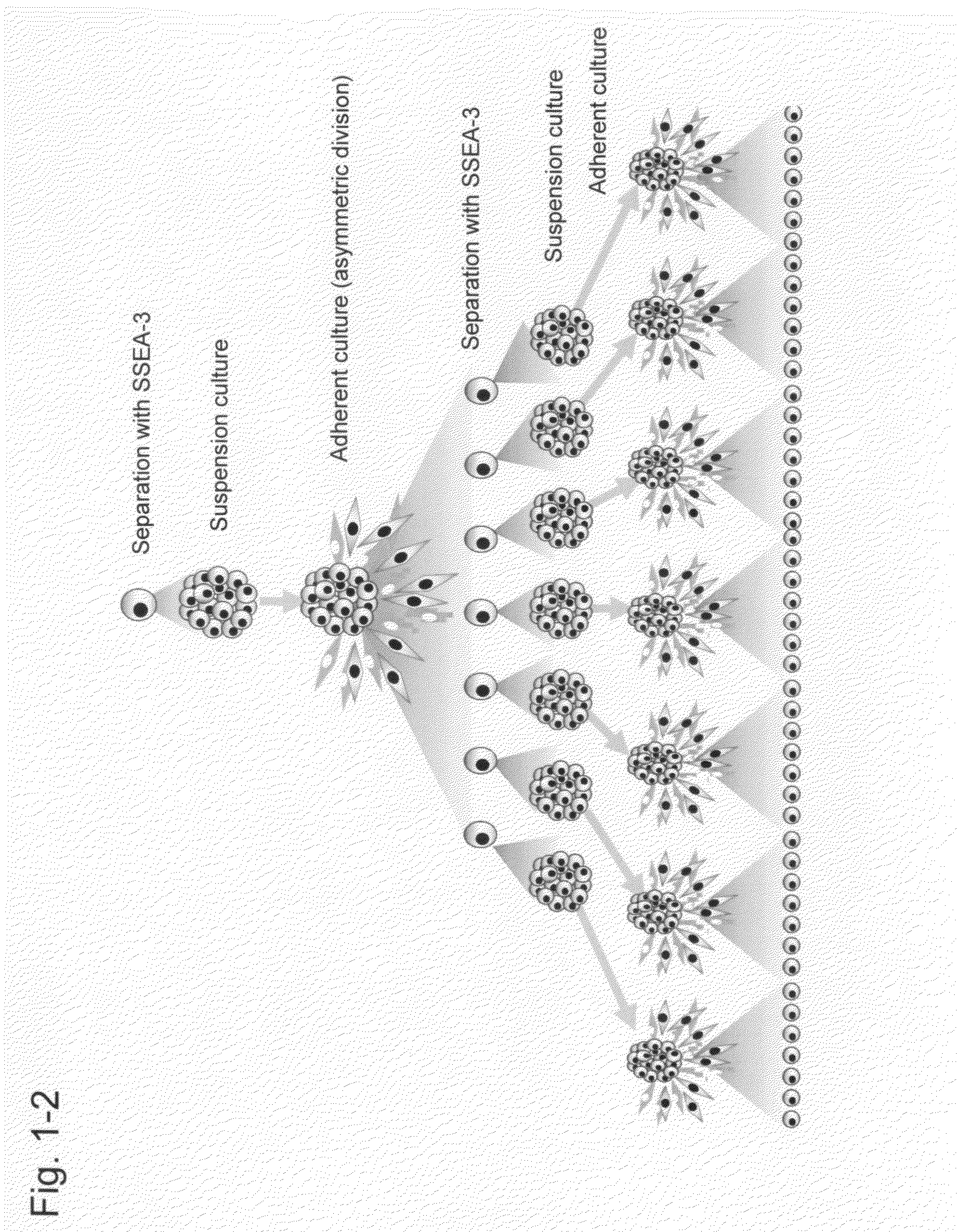
The pluripotent stem cells of the present invention are capable of differentiating into the three germ layers through in vitro adherent culture. Specifically, the pluripotent stem cells can differentiate into cells representative of the three germ layers, skin, liver, nerve, muscle, bone, fat, and the like through in vitro induction culture. Also, the pluripotent stem cells are capable of differentiating into cells characteristic of the three germ layers when transplanted in vivo into the testis. Furthermore, the pluripotent stem cells are capable of surviving and differentiating into organs (e.g., skin, spinal cord, liver, and muscle) when transplanted to the damaged organs via intravenous injection into a living body.

Problems solved by technology

However, in the case of higher organisms such as humans, the tissue regenerative capacity is far lower than that of those animals.
However, such pluripotency becomes limited as development proceeds, followed by cell differentiation for specialization resulting in each type of tissue.
However, it has remained unknown whether or not pluripotent stem cells are present in mature mammalian bodies, as in the case of planarians or newts.

Method used


Image

  • Pluripotent stem cell that can be isolated from body tissue
  • Pluripotent stem cell that can be isolated from body tissue
  • Pluripotent stem cell that can be isolated from body tissue

Examples


example 1

Preparation and Characterization of Muse-Enriched Cell Fractions and M-Clusters

Materials and Methods

The following cells are used in Examples.

Two strains of human dermal fibroblast fractions (H-fibroblasts) and four strains of human MSC (bone marrow stromal cell) fractions (H-MSC fractions) were used as mesenchymal cells. Human fibroblast fractions were (1) H-fibroblast-1 (normal human fibroblast cells (NHDF), Lonza), and (2) H-fibroblast-2 (adult human dermal fibroblasts (HDFA, ScienCell, Carlsbad, Calif.)). Human MSC fractions, H-MSC-1, -2 and -3 were obtained from Lonza, and H-MSC-4 was obtained from ALLCELLS. Human MSC fractions are specifically described in Pittenger, M. F. et al. Science 284, 143-147 (1999); Dezawa, M. et al. J Clin Invest 113, 1701-1710 (2004); and Dezawa, M. et al. Science 309, 314-317 (2005).

Cells were cultured at 37° C. in α-MEM (alpha-minimum essential medium) containing 10% FBS and 0.1 mg / ml kanamycin with 5% CO2. Cells cultured directly after their shipm...

example 2

Characterization of Muse Cells Isolated Using SSEA-3

Examination in Example 1 revealed that SSEA-3 (+) cell fractions obtained by FACS had the properties of pluripotent stem cells; that is, they were Muse cells (J, K, and the like above). Furthermore, in vitro differentiation ability and in vivo differentiation ability were examined using isolated SSEA-3 (+) cells and then Muse-derived iPS cells were established.

1. Examination of In Vivo Differentiation Ability by Transplantation of Cells into Damaged Tissues

SSEA-3 (+) Muse cells labeled with GFP (green fluorescent protein)-lentivirus were isolated and then transplanted via intravenous injection into immunodeficient mice (NOG mice) with damaged spinal cord (Crush injury of spinal cord), damaged liver (intraperitoneal injection of CCl4, fulminant hepatitis model), or damaged gastrocnemius (muscle) (cardiotoxin injection). Human skin cell-derived Muse cells were labeled with GFP (green fluorescent protein)-lentivirus (Hayase M et al., ...


PUM


Abstract

Objects of the present invention are to provide a method for directly obtaining pluripotent stem cells from body tissue and the thus obtained pluripotent stem cells. The present invention relates to SSEA-3 (+) pluripotent stem cells that can be isolated from body tissue.

Description

BACKGROUND OF THE INVENTION1. Field of the InventionThe present invention relates to body tissue-derived Pluripotent stem cells.2. Background ArtPlanarians and newts can regenerate their entire bodies even after their bodies have been cut. Such high regenerative capacity depends on the presence of pluripotent stem cells existing in the mesenchymal tissues. However, in the case of higher organisms such as humans, the tissue regenerative capacity is far lower than that of those animals. An inner cell cluster (or ICM: inner cell mass) in a mammalian blastocyst is recognized as a collection of pluripotent stem cells that is capable of differentiating into cells of ectodermal, mesodermal, and endodermal cell lineages. However, such pluripotency becomes limited as development proceeds, followed by cell differentiation for specialization resulting in each type of tissue.In recent years, adult stem cells or tissue stem cells that can contribute to tissue regeneration have been attracting at...

Claims


Application Information

Patent Timeline
IPC IPC(8): C12N5/071
CPCC12N5/0607C12N2509/00C12N5/0696C12N2506/1353A61P1/00A61P1/04A61P1/16A61P1/18A61P13/00A61P13/10A61P13/12A61P15/00A61P17/00A61P17/02A61P19/00A61P21/00A61P25/00A61P27/02A61P3/00A61P35/00A61P37/02A61P43/00A61P7/00A61P9/00A61K35/12C12N15/1138C12N5/0662C12N5/0663C12N15/86C12N5/0605C12N5/0667C12N5/0668A61K35/50A61K35/28
Inventor DEZAWA, MARIFUJIYOSHI, YOSHINORINABESHIMA, YOUICHIWAKAO, SHOHEI
Owner DEZAWA MARI
Learn More