
Rapid Detection of Post-Vaccination Antibody Response

a technology of antibody response and detection method, which is applied in the field of rapid detection of post-vaccination antibody response, can solve the problems of rare and time-consuming monitoring of the efficacy of administered vaccines, and achieve the effect of reducing the number of false positives

Inactive Publication Date: 2010-12-23
SURAPANENI KRISHNA P +3
View PDF8 Cites 16 Cited by
  • Summary
  • Abstract
  • Description
  • Claims
  • Application Information

AI Technical Summary

Problems solved by technology

The administered vaccines are, however, rarely monitored for efficacy.
Such studies employ ELISA or Neutralization tests, however, are time consuming and only cover a few vaccine subjects.

Method used


Image

  • Rapid Detection of Post-Vaccination Antibody Response
  • Rapid Detection of Post-Vaccination Antibody Response
  • Rapid Detection of Post-Vaccination Antibody Response

Examples


second embodiment

[0050]In another embodiment, the present invention is directed towards a rapid detection kit that employs a lateral flow technique to simultaneously screen for the presence of a plurality of specific antibodies reactive with analytes such as bacteria, viral proteins and allergens in immunized individuals. Thus, in a second embodiment, the present invention employs an improved lateral flow method by arranging several lateral flow membranes radially with a single central sample application pad.

[0051]The method and device of the present invention, in one embodiment, detects IgG antibodies in the serum and IgA in fecal matter and saliva (or other human body fluids). In one embodiment, antigens are immobilized on a test membrane strip, such as nitrocellulose. The antibodies present in body fluids form a complex with colloidal gold / colored latex particles. Once the complex is formed, it moves across a series of membranes, and produces a colored line of antigen-antibody complex to indicate...

first embodiment

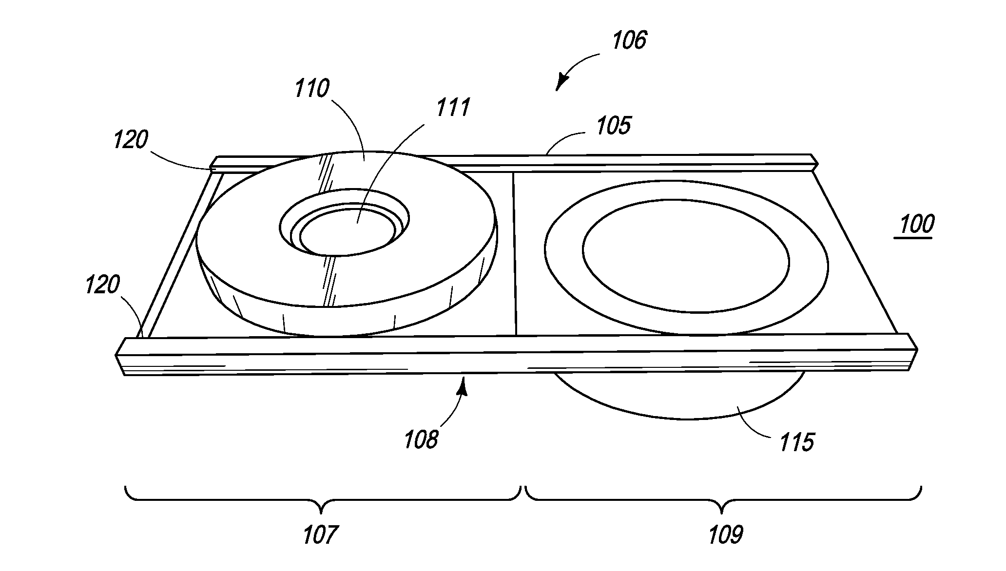
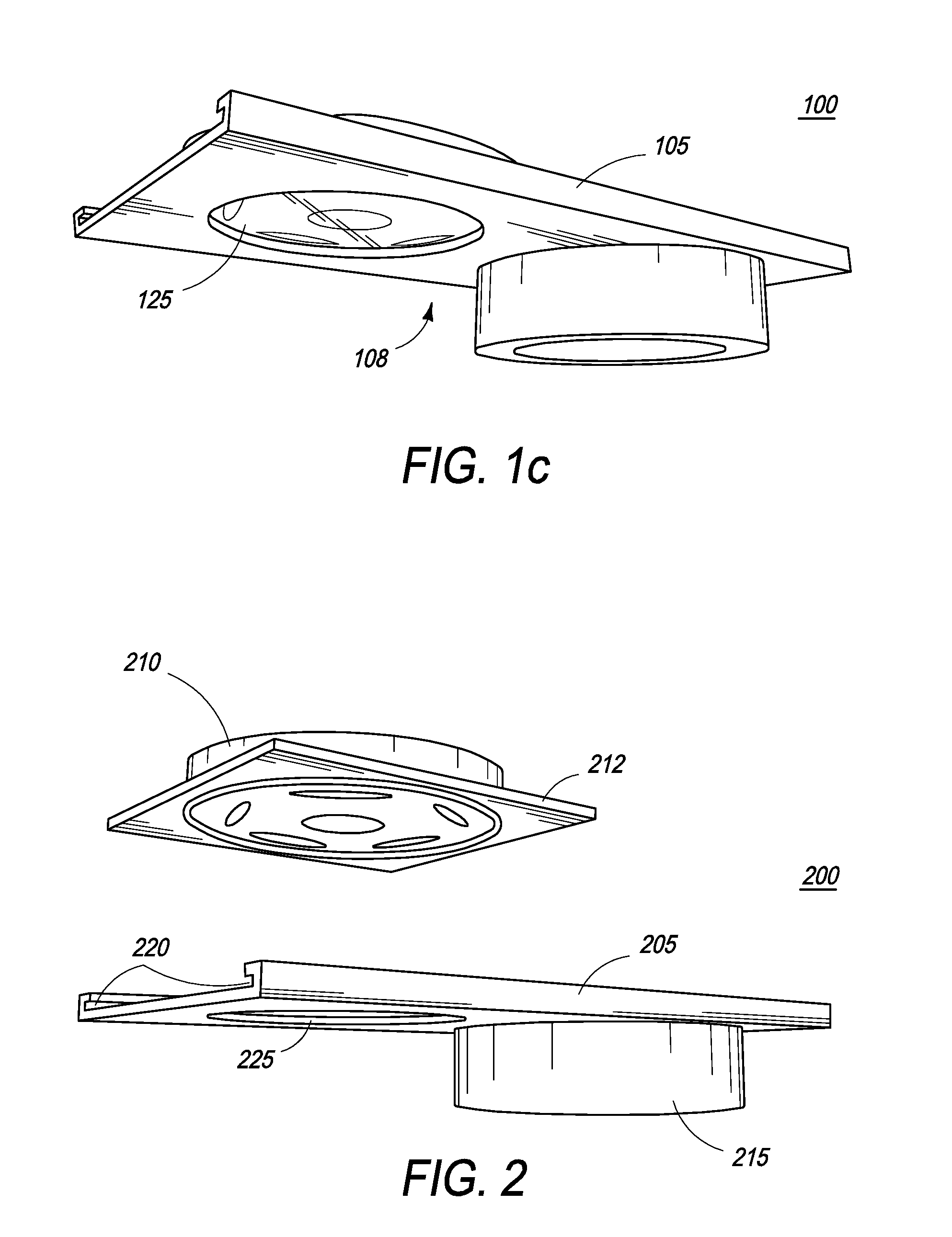
[0057]In a first embodiment, the nitrocellulose membrane is contained within and integral to a removable pillbox container, having various layers, including the sample application pad, conjugate pad, and other membranes.

[0058]In a second embodiment, the nitrocellulose membrane is part of a support platform and is separated from the other membranes by employing a mesh screen, comprised of a material such as nylon, as part of the pillbox container, so that the membrane can be viewed by sliding away the various other membranes.

[0059]The intensity of the reaction can be quantified by a conventional hand-held colorimeter. Thus, in one embodiment, the rapid detection test kit of the present invention provides a quantitative as well as qualitative in vitro diagnostic for the detection of antibodies to viruses and / or bacteria in human body fluids and thus is very effective in qualifying the success of vaccinations.

[0060]The present invention is directed toward multiple embodiments. Referenc...


PUM

PropertyMeasurementUnit
sizeaaaaaaaaaa
sizeaaaaaaaaaa
temperaturesaaaaaaaaaa

Abstract

The present inventions are directed to apparatuses for rapidly measuring post-vaccination immune status. In one version, the apparatus has a support platform, with a top side, a bottom side, a first portion, and a second portion. A first void is integrally formed in the first portion. A container is configured to be removably affixed to the top side of the support platform. The container has a housing, a base, and at least one reactant. The container base can be viewed through the first void when the container is removably affixed to the first portion of the top side. An absorbent material is affixed to the second portion, where the base of the container comes into contact with the absorbent material when the container is removably affixed to the second portion of the top side.

Description

CROSS-REFERENCE[0001]The present application relies on U.S. Patent Provisional Application No. 61 / 058,866, which was filed on Jun. 4, 2008.FIELD OF THE INVENTION[0002]The present invention generally relates to apparatuses, methods, and compositions for detecting the presence of antibodies in body fluids, including blood, serum, plasma, tears, lactate, saliva, urine or feces. The present invention also relates generally to apparatuses, methods, and compositions for detecting the presence of antibodies in body fluids to determine the immune status of vaccinated individuals by using various detectable analytes, such as bacteria, protozoa, viral proteins, allergens, viral cell lysates, bacterial cell lysates, and carbohydrates as capture antigens. The present invention relates, more specifically, to apparatuses, methods, and compositions for detecting the presence of antibodies in human body fluids by using a sink-in (hereinafter referred to as “SINK-SORB”) technology.BACKGROUND OF THE ...

Claims


Application Information

Patent Timeline
Patent Type & Authority Applications(United States)
IPC IPC(8): G01N30/00
CPCG01N2333/105G01N33/558G01N33/54388G01N33/54391
Inventor SURAPANENI, KRISHNA P.GANDLUR, PRASADARAOSURYANARAYANA, VEDNLA VENKATAPATHAK, AJAI KUMAR
Owner SURAPANENI KRISHNA P
Learn More