
Ultrasound system and method to automatically identify and treat adipose tissue

a technology of ultrasound and adipose tissue, applied in the field of diagnostic imaging and therapy systems, can solve the problems of system invasiveness, lack of control of localization of hifu system, and inability to provide visual representation or image of volume, etc., and achieve the effect of facilitating the user of the system

Inactive Publication Date: 2010-11-11
GENERAL ELECTRIC CO
View PDF100 Cites 54 Cited by
  • Summary
  • Abstract
  • Description
  • Claims
  • Application Information

AI Technical Summary

Benefits of technology

[0006]Accordingly, there is a need for ultrasound imaging and therapy systems that indicate where, within a volume of the patient, therapy has been provided or will be provided. Furthe

Problems solved by technology

Some systems may be invasive, such as liposuction, where a device is inserted into the body and physically removes adipose tissue through suction.
However, since the ultrasound signals may have a harmful effect on the non-adipose tissue, it is important for a user of a HIFU system to know and control where treatment has been provided within the body of a patient.
For example, the HIFU system may only display the surface of the patient's skin and does not provide a visual representation or image of the volume of the body under the surface.
Consequently, the above HIFU system does not provide control for localizing therapy to certain regions under the surface of the skin.
Further, the above conventional HIFU system also does not know or determine where non-adipose tissue may be located with respect to the adipose tissue.
Therefore, the HIFU system does not automatically differentiate the tissues.
The HIFU system may also not confirm that therapy has been delivered to the desired regions.

Method used


Image

  • Ultrasound system and method to automatically identify and treat adipose tissue
  • Ultrasound system and method to automatically identify and treat adipose tissue
  • Ultrasound system and method to automatically identify and treat adipose tissue

Examples


Embodiment Construction

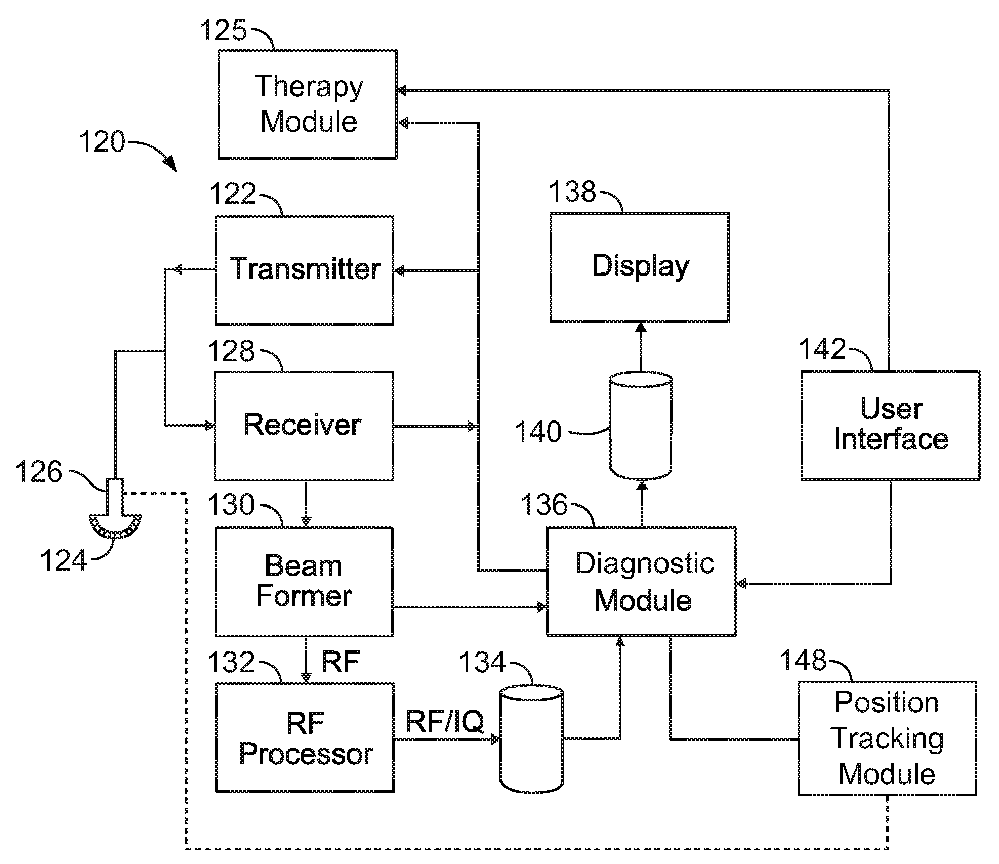
[0024]Exemplary embodiments that are described in detail below include ultrasound systems and methods for imaging and treating a region of interest (ROI). The ROI may include adipose tissue and / or non-adipose tissue, such as muscle tissue, bone, tissue of organs, and blood vessels. The system may display the ROI so that an operator or user of the system can distinguish the adipose tissue and the non-adipose tissue and / or the system may automatically differentiate the adipose tissue and the non-adipose tissue prior to treating. Treatment of the ROI may include providing high-intensity focused ultrasound (HIFU) signals to treatment locations within the ROI. For example, HIFU signals may be directed to treatment locations within the adipose tissue to at least partially liquefy the adipose tissue. Liquefication may occur through cell lysis, cavitation, and / or thermal damage in the adipose tissue.

[0025]The following detailed description of certain embodiments will be better understood wh...


PUM


Abstract

An ultrasound imaging and therapy system that includes an ultrasound probe and an ultrasound diagnostic module to control the probe to obtain diagnostic ultrasound signals from a region of interest (ROI). The ROI includes adipose tissue and non-adipose tissue. The diagnostic module analyzes the diagnostic ultrasound signals and automatically differentiates adipose tissue from non-adipose tissue. The system also includes an ultrasound therapy module to control the probe to deliver, during a therapy session, a therapy at a treatment location based on a therapy parameter to the adipose tissue differentiated by the ultrasound diagnostic module. A method for delivering therapy to a region of interest (ROI) in a patient is also provided.

Description

CROSS-REFERENCE TO RELATED APPLICATIONS[0001]This application includes subject matter that is similar to the subject matter described in U.S. Patent Applications having Attorney Docket No. 235594 (555-0003US), entitled “ULTRASOUND SYSTEM AND METHOD TO DELIVER THERAPY BASED ON USER DEFINED TREATMENT SPACES,” and Attorney Docket No. 235610 (555-0005US), entitled “ULTRASOUND SYSTEM AND METHOD TO DETERMINE MECHANICAL PROPERTIES OF A TARGET REGION,” both of which are filed contemporaneously herewith and are incorporated by reference in their entirety.BACKGROUND OF THE INVENTION[0002]The subject matter herein relates generally to diagnostic imaging and therapy systems that provide diagnostic imaging and treatment of a region of interest in a patient, and more particularly, to ultrasound systems that image and treat adipose tissue.[0003]Various body contouring systems exist today that attempt to remove or destroy fatty tissue (or adipose tissue) from a person's body. Some systems may be in...

Claims


Application Information

Patent Timeline
IPC IPC(8): A61N7/00A61B8/13
CPCA61B8/08A61B8/485A61B8/0841A61B8/0858A61B8/14A61B8/4405A61B8/4411A61B8/4427A61B8/4455A61B8/4483A61B8/463A61B8/467A61B8/469A61B8/483A61B19/201A61B19/56A61B2019/5276A61N7/02A61N2007/0008A61B8/0833A61B8/461A61B8/4245A61B34/25A61B90/11A61B2090/378
Inventor LEE, WARRENARORA, DHIRAJLANDBERG DAVIS, CYNTHIA ELIZABETHFAN, YINGHAZARD, CHRISTOPHER ROBERTRIGBY, KENNETH WAYNESMITH, LOWELL SCOTTTHOMENIUS, KAI ERIK
Owner GENERAL ELECTRIC CO
Learn More