Methods for measuring cell response to DNA damaging agents using promyelocytic leukemia protein nuclear bodies
a technology cell response, which is applied in the field of cell response monitoring of dna damage agent using promyelocytic leukemia protein nuclear body, can solve the problems of unsatisfactory tumour volume as a marker for treatment efficacy, unable to fully understand how the nuclear activity contributes to the nuclear activity, etc., and achieves the effect of altering the balance of forces constraining chromatin
- Summary
- Abstract
- Description
- Claims
- Application Information
AI Technical Summary
Benefits of technology
Problems solved by technology
Method used
Image
Examples
example 1
The Increase in PML NB Number in Response to DSBs is Sensitive, Rapid and Dose-Dependent
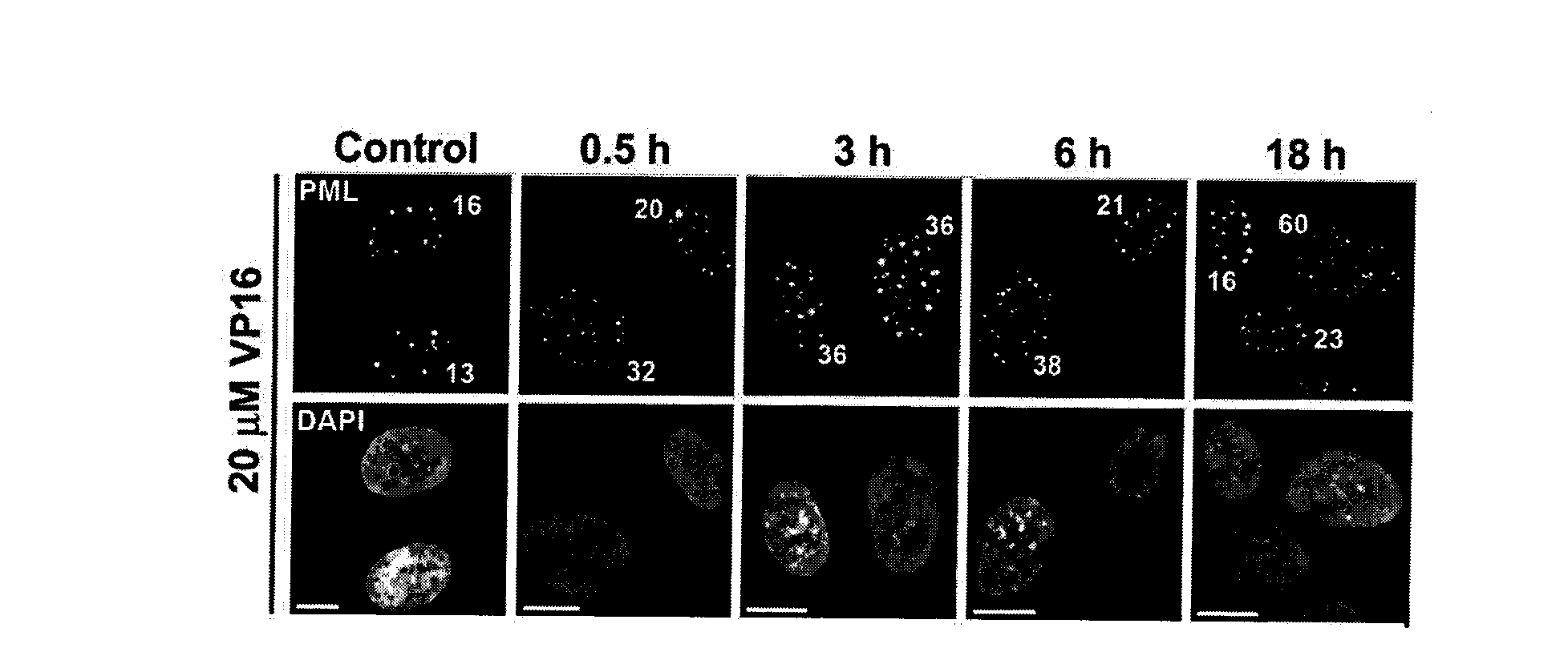
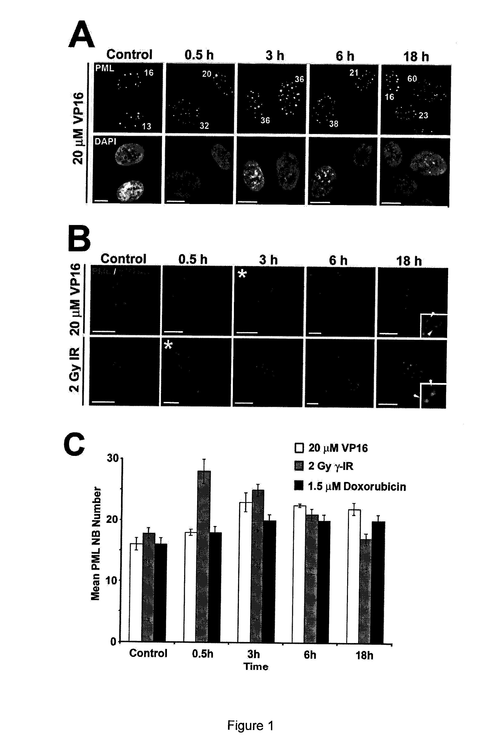
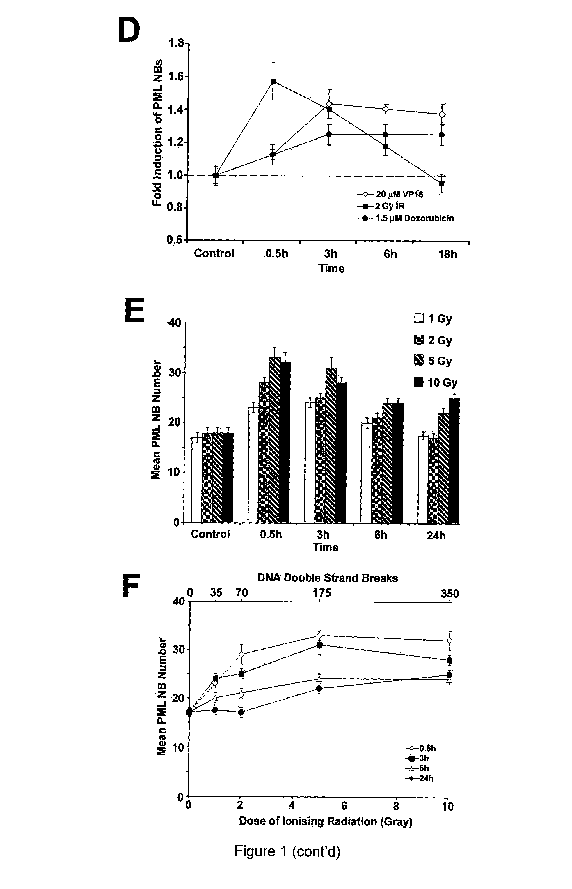
[0116]The response of PML NBs to DSBs in a normal human diploid fibroblast (NHDF) cell line GM05757 was assessed, using ionising radiation (IR), etoposide (VP16) and doxorubicin (FIG. 1). IR generates both single-strand breaks (SSBs) and DSBs in DNA, whereas, the topoisomerase II-inhibitors VP16 and doxorubicin create primarily DSBs (reviewed by Kurz and Lees-Miller, 2004). PML NBs were counted in maximum intensity Z-projections of individual cells. PML NB number increased following DSB induction (FIG. 1A). Furthermore, we found that the time point associated with the highest number of PML NBs coincided with the peak of H2AX phosphorylation (γ-H2AX; FIG. 1B), an event that occurs on chromatin surrounding DSBs (Rogakou et al., 1999). Maximum PML NB number correlated with peak γ-H2AX signal regardless of the method of DSB-induction, suggesting that the increase in PML NB number is coupled to DSB fo...
example 2
PML Protein and NB Dynamics in Response to DSBs
[0118]The dynamics of PML NBs following DSB induction with VP16 was examined by live cell analysis of U-2 OS cells stably expressing PML isoform IV (FIG. 2). We found that within 5 min after addition of VP16, new and smaller PML-containing structures began to appear adjacent to the larger PML NBs that were present before treatment (FIG. 2A). These new bodies, which we term microbodies, arise from pre-existing PML NBs by a supramolecular fission mechanism, as confirmed by spinning-disc confocal microscopy (FIG. 2B). This fission mechanism is similar to that observed for new PML NB formation in early S-phase (Dellaire et al., 2006), as PML NB biochemical composition was initially indistinguishable between microbodies and the larger parental PML NBs with respect to Sp 100 and SUMO-1 content (FIG. 11A-B). However, although Sp100 levels at PML NBs did not change over the time course observed (FIG. 11A), we did notice a reproducible drop in S...
example 3
Structural Destabilization of PML NBs Correlates Closely with Topological Changes in Chromatin Following DNA Damage
[0121]To address the ultrastructural changes in PML NBs following DNA damage, we employed immuno-gold detection of PML with correlative light and electron spectroscopic imaging (LM / ESI, Dellaire et al., 2004)(FIG. 4). Using LM / ESI we observed that in control NHDFs, PML NBs exhibit radial symmetry and make extensive contacts with the surrounding chromatin (FIG. 4A). Upon treatment with VP16, we found that PML NBs lose their radial symmetry and make fewer contacts with the surrounding chromatin fibres. We also observed “microbody-like” structures, identified by immunogold detection of PML, adjacent to chromatin in the vicinity of larger “parental” PML NBs (FIG. 4B-C). A much larger interchromatin domain (ICD) space was also apparent in cells treated with VP16 (i.e. black spaces outside of chromatin in FIG. 4). These changes in both chromatin and PML NBs are reminiscent of...
PUM
| Property | Measurement | Unit |
|---|---|---|
| Stress optical coefficient | aaaaa | aaaaa |
| Morphology | aaaaa | aaaaa |
Abstract
Description
Claims
Application Information
 - R&D
- Intellectual Property
- Life Sciences
- Materials
- Tech Scout
- Unparalleled Data Quality
- Higher Quality Content
- 60% Fewer Hallucinations
Browse by: Latest US Patents, China's latest patents, Technical Efficacy Thesaurus, Application Domain, Technology Topic, Popular Technical Reports.
© 2025 PatSnap. All rights reserved.Legal|Privacy policy|Modern Slavery Act Transparency Statement|Sitemap|About US| Contact US: help@patsnap.com


