
Ultrasound diagnosis apparatus and a centesis supporting method

a technology of ultrasonic diagnosis and supporting method, which is applied in the direction of fluid coupling, application, tomography, etc., can solve the problems of inability to accurately determine the position or direction of the needle, and the needle is inserted in an unexpected direction, so as to achieve safe and efficient examination or medical, the effect of easy determination

Active Publication Date: 2010-09-23
TOSHIBA MEDICAL SYST CORP
View PDF3 Cites 24 Cited by
  • Summary
  • Abstract
  • Description
  • Claims
  • Application Information

AI Technical Summary

Benefits of technology

[0010]The present invention addresses the above-mentioned and other problems and drawbacks, in order to provide a novel ultrasound diagnosis apparatus and a centesis supporting method that can accurately and easily determine a relative misalignment of a centesis needle to a target region, while the centesis needle inserted into a patient's body travels in an unexpected direction due to unevenness of anatomy hardness.
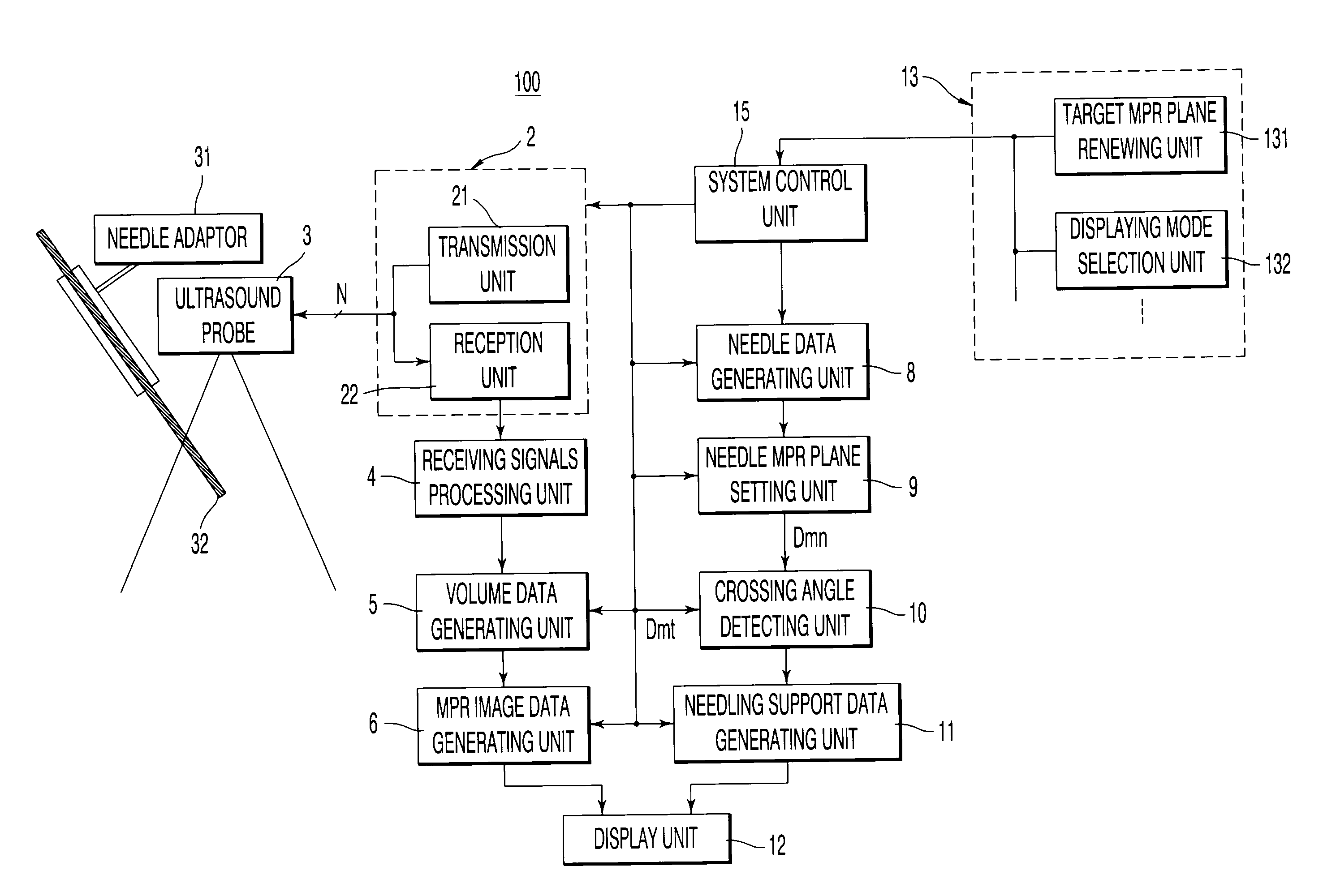
[0035]According to the present invention, it becomes possible to display a cross-sectional plane including a target set up by an operator and a scheduled pathway of a centesis needle overlapped with a centesis needle tracking plane generated by automatically detecting the centesis needle or overlapped with a projected image of the centesis needle. Consequently, it becomes possible to safely and efficiently perform an examination or medical treatments, even when a centesis needle inserted into a patient body travels in an unexpected direction, since it becomes possible to visually accurately and easily determine a relative misalignment of the centesis needle to a target region.

Problems solved by technology

However, in reality it actually happens that the needle is inserted in an unexpected direction due to unevenness of living body hardness along the predetermined inserting pathway.
Further, according to the conventional position detecting method using 3D image data, it also becomes impossible to accurately determines the position or the direction of the needle inserted into a body, since the needle is inserted in a different direction from the predetermined inserting pathway indicated on the image data detected by a position sensor.

Method used


Image

  • Ultrasound diagnosis apparatus and a centesis supporting method
  • Ultrasound diagnosis apparatus and a centesis supporting method
  • Ultrasound diagnosis apparatus and a centesis supporting method

Examples


Embodiment Construction

[0055]An ultrasound diagnosis apparatus consistent with the present invention firstly sets up a first multi-planar-reconstruction (MPR) cross-sectional plane (hereinafter, simply referred to as a first “MPR plane”) including the target region. The first MPR plane is referred to as a target MPR plane. Further, the ultrasound diagnosis apparatus generates a target image data at the target MPR plane of volume data acquired through a 3D scan during the needle insertion time, and also generates centesis needle data indicating positions and directions of the needle based on the volume data. Then, the ultrasound diagnosis apparatus detects a crossing angle between a second MPR plane including the needle data (i.e., a centesis needle MPR plane) and the target MPR plane, and generates a second MPR image data (needle image data) by setting up the needle MPR plane on the volume data during the needle insertion time. The centesis support data is generated by composing the target image data and ...


PUM


Abstract

An ultrasound diagnosis system and a centesis support controlling method by which positions and directions of a centesis needle traveling to a target region can accurately and easily be determined, including an MPR image data generating unit which generates target image data by setting a prescribed target MPR plane on volume data acquired through 3D scan over a 3D volume including a target region; a centesis needle MPR plane setting unit which generates centesis needle data indicating position and directions of the centesis needle, based on the acquired volume data; a crossing angle detection unit which detects a crossing angle between the target MPR plane and the centesis needle MPR plane including the centesis needle data; and a needling support data generating unit which generates needling support data by composing the target image data and the centesis needle data overlapped with centesis needle image data based on the detected crossin angle.

Description

[0001]This application claims priority under 35 U.S.C. §119(a) from, and the benefit of, Japanese Patent Application No. 2009-70793 filed Mar. 23, 2009, the contents of which are expressly incorporated herein by reference in their entirety.BACKGROUND OF THE INVENTION[0002]A. Field of the Invention[0003]The present invention relates to an ultrasound diagnosis apparatus and a centesis supporting method, and more particularly, to an ultrasound diagnosis apparatus and a centesis supporting method that can visually recognize a centesis needle insertion status into a target region in a patient by automatically tracking the inserted needle while observing ultrasound image data.[0004]B. Background of the Invention[0005]An ultrasound diagnosis apparatus transmits and receives ultrasound through a plurality of ultrasound transducers installed in a tip portion of an ultrasound probe to and from a diagnostic target region in an object. By simply touching an ultrasound probe to the surface of a ...

Claims


Application Information

Patent Timeline
Patent Type & Authority Applications(United States)
IPC IPC(8): A61B8/14
CPCA61B8/00A61B17/3403A61B2017/3413A61B8/483A61B2019/507A61B2019/5236A61B2019/5289A61B2019/4836A61B2090/364A61B2090/0807A61B2034/107A61B2090/374
Inventor ICHIOKA, KENICHISUMI, ATSUSHIKAKEE, AKIHIRO
Owner TOSHIBA MEDICAL SYST CORP
Learn More