
Storage system for storing the sampling data of pathological section and method thereof

a storage system and pathological section technology, applied in the field of storage system for storing pathological section sampling data, can solve the problems of large storage space, large amount of manpower, capital investment in archiving process, etc., and achieve the effect of fast biopsy analysis

Inactive Publication Date: 2010-07-01
EBM TECH
View PDF11 Cites 4 Cited by
  • Summary
  • Abstract
  • Description
  • Claims
  • Application Information

AI Technical Summary

Benefits of technology

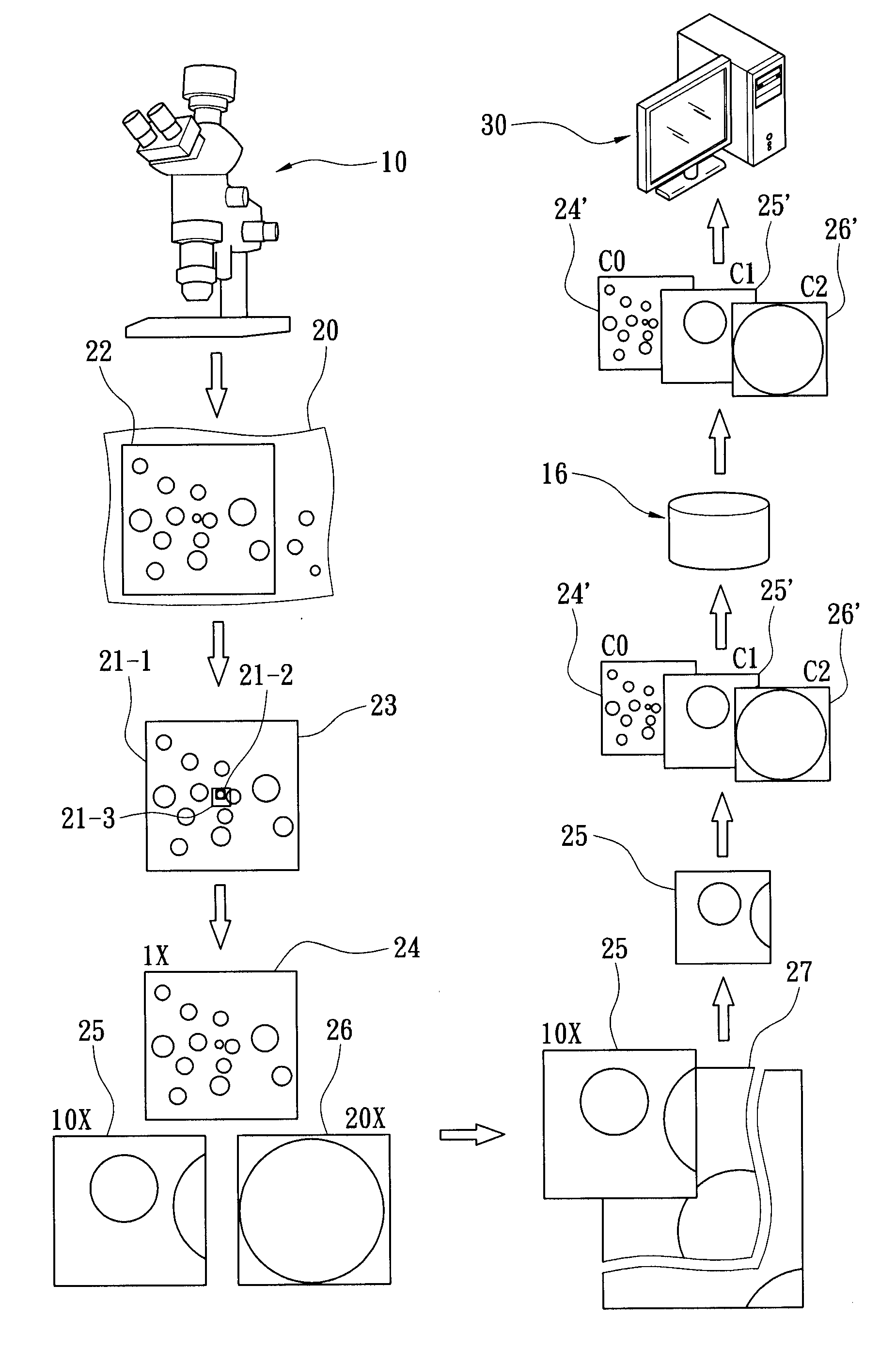
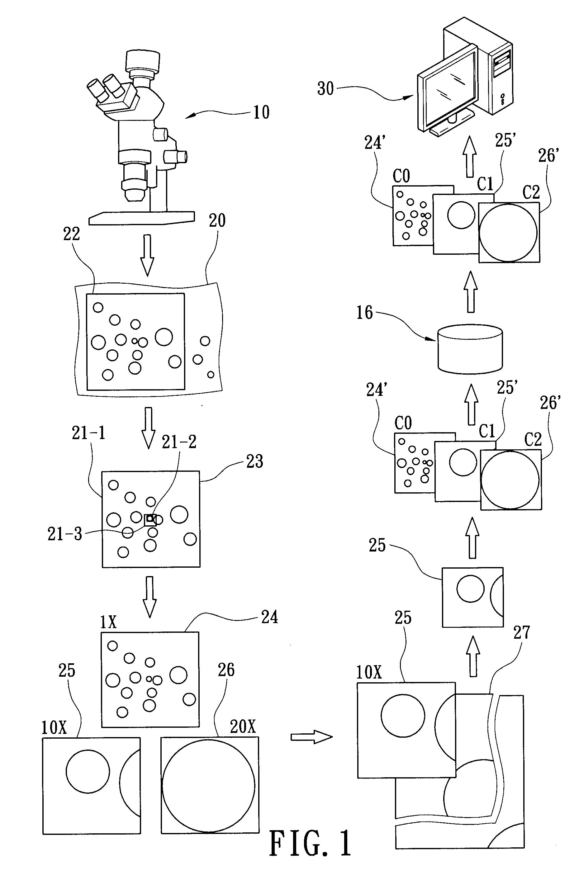
[0008]In view of the aforementioned issues, the present invention provides a storage system for sampling data of pathological section and method thereof, which takes variously magnified tissue images from the pathological section, compares respectively each of the variously magnified tissue images with the original image enlarged to the same degree of magnification (the original symptom image enlarged with the same magnification as the tissue image currently being compared, a.k.a. original image with matching magnification), and generates the coordinates of the variously magnified tissue images corresponding to the original symptom image, then integrates and stores the variously magnified tissue images and their corresponding coordinates into a sampling data (i.e. the sampling data of pathological sections), which allows physicians to explicitly appreciate the position of the magnified tissue images in the original symptom image when viewing the tissue images, facilitating fast biopsy analysis.
[0011]In summary, the storage system for storing sampling data of pathological section and the storage method thereof according to the present invention compares variously magnified tissue images against the original symptom image with matching magnification to acquire the corresponding image coordinates of the variously magnified tissue image on the original symptom image, then integrates the variously magnified tissue image and the corresponding image coordinate into the sampling data. In this way, when physicians are viewing the magnified tissue images, they can explicitly appreciate the relative positions of variously magnified tissue images on the original symptom image through simple search operations, further providing rapid and correct biopsy analysis, thus improving medical treatment quality.

Problems solved by technology

Conventionally, medical data of patients are displayed on physical documents by doctors (e.g. medical history data in the form of papers and file folders, X-ray films and the like), which occupy large storage space and consume huge amount of resources such as manpower, capital investment for archiving processes.
Many medical instruments offer digitalized medical data, but still large portion therein are not stored in standardized digital formats.
However, after enlargement of tissue image, in case of lacking relevant supportive information such as relative coordinate, enlargement multiplicities and so on, it becomes meaningless graphics, thus impossible to revert to original state as understandable tissue image information.

Method used


Image

  • Storage system for storing the sampling data of pathological section and method thereof
  • Storage system for storing the sampling data of pathological section and method thereof
  • Storage system for storing the sampling data of pathological section and method thereof

Examples


Embodiment Construction

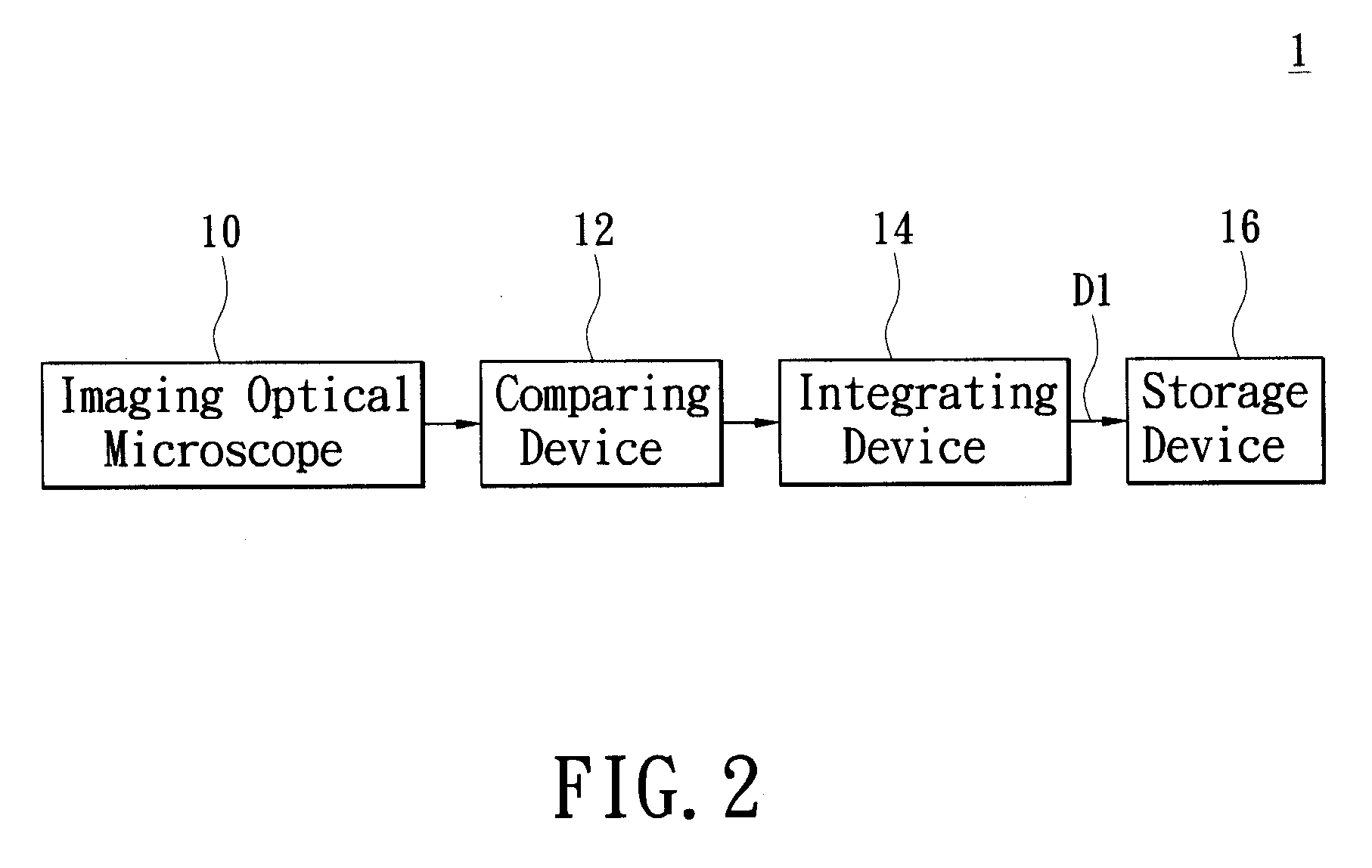
[0017]Refer now to FIG. 1 wherein a flowchart of the storage method for sampling data of pathological section (i.e. biopsy) according to the present invention is shown. Meanwhile, refer also to FIGS. 3 and 4, in which a flowchart of the method according to the present invention is respectively shown. First of all, select an imaging microscope 10, then place the pathological section 20 obtained from the body of a patient on the observation stage of the imaging microscope 10 (S100), wherein the pathological section 20 is a test section for microscope observation made from pathological tissue such as tissue section of human tissue blood, bacteria, or excrement. Then, select an observation region on the pathological section 20, and use the imaging microscope 10 to capture an image of the selected observation region in order to generate an original symptom image 23 (S102). Next, select a region of interest (ROI) 21-1 from the generated original symptom image 23 (S104), and then enlarge t...


PUM


Abstract

A storage system and method thereof for storing the sampling data of a pathological section, the steps of the method includes taking variously magnified tissue images from the pathological section; moreover, comparing respectively the variously magnified tissue images with an original image with matching magnification so as to generate the coordinates of the variously magnified tissue images corresponded to the original symptom image; furthermore, integrating the variously magnified tissue images and its corresponded coordinates into a sampling data, then storing the sampling data.

Description

BACKGROUND OF THE INVENTION[0001]1. Field of the Invention[0002]The present invention relates to a storage system for storing sampling data of pathological section and method thereof; in particular, to a storage system and method thereof for improving medical data management and storage.[0003]2. Description of Related Art[0004]As computer technologies advance, digitalization of medical information and management of high efficiency thereof have now become important and necessary trends. Conventionally, medical data of patients are displayed on physical documents by doctors (e.g. medical history data in the form of papers and file folders, X-ray films and the like), which occupy large storage space and consume huge amount of resources such as manpower, capital investment for archiving processes. Presently, it is possible to use computer technologies to perform systematic management on medical data by means of database, in conjunction with data transfer over computer network, allowing ...

Claims


Application Information

Patent Timeline
Patent Type & Authority Applications(United States)
IPC IPC(8): G06K9/00G16H10/40G16H30/40
CPCG06K9/00134G06F19/321G16H10/40G16H30/40G06V20/693
Inventor PAN, WILLIAM
Owner EBM TECH
Learn More