Cancer diagnostic method and system

a diagnostic method and system technology, applied in the field of cancer diagnostic methods and systems, can solve the problems of previous researchers not yet producing a difficult to classify malignant and benign cancers, and difficult to achieve the effect of achieving stable and accurate system

Active Publication Date: 2010-05-13
ZHANG PING
View PDF18 Cites 17 Cited by
  • Summary
  • Abstract
  • Description
  • Claims
  • Application Information

AI Technical Summary

Problems solved by technology

Current image processing techniques make breast abnormality detection easier, however classification of malignant and benign cancers is still a very challenging and a difficult problem for researchers.
However, previous researchers have not yet produced a stable and accurate system, and classification of malignant and benign cancers is still a very challenging problem for researchers.

Method used


Image

  • Cancer diagnostic method and system
  • Cancer diagnostic method and system
  • Cancer diagnostic method and system

Examples


Embodiment Construction

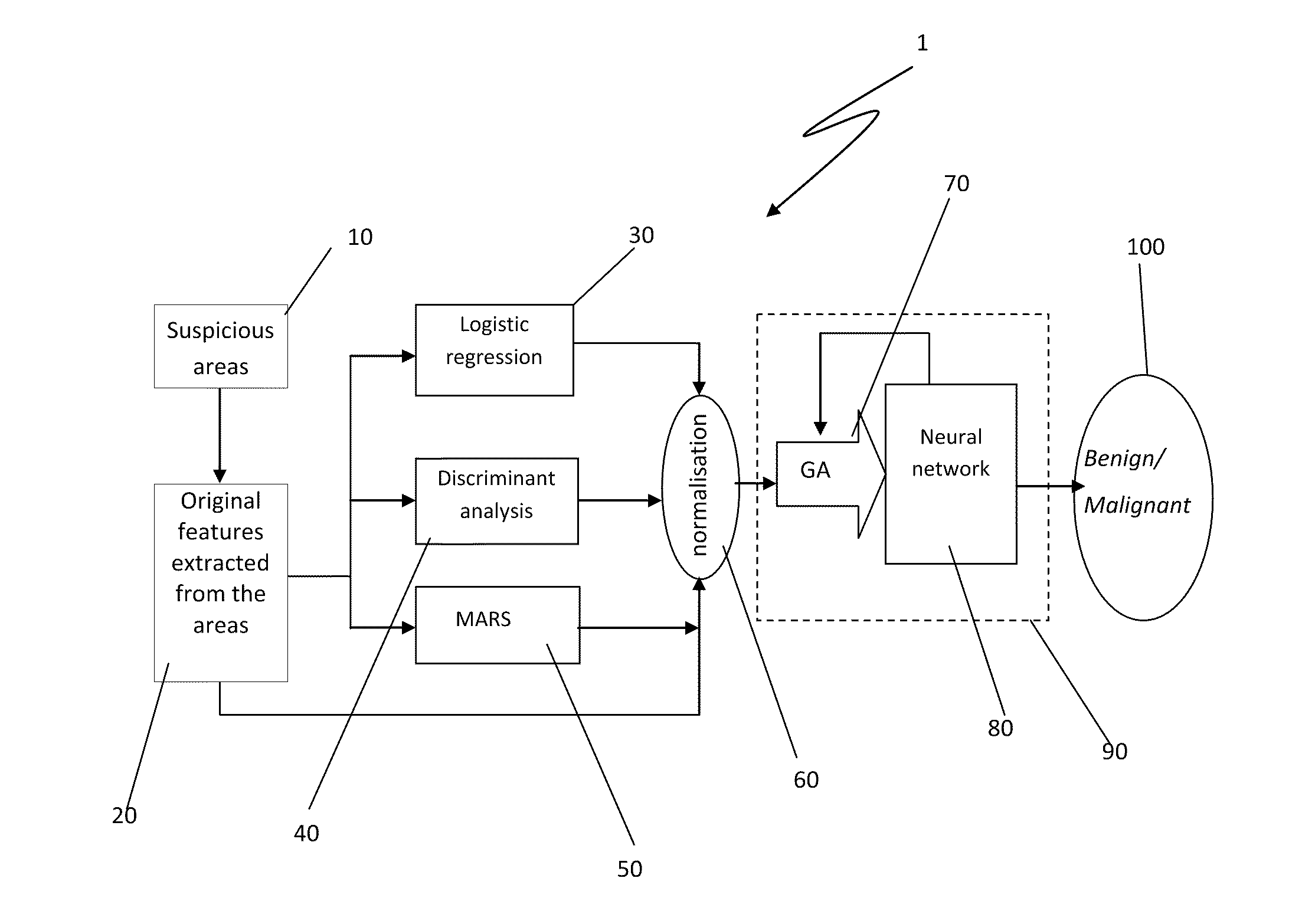
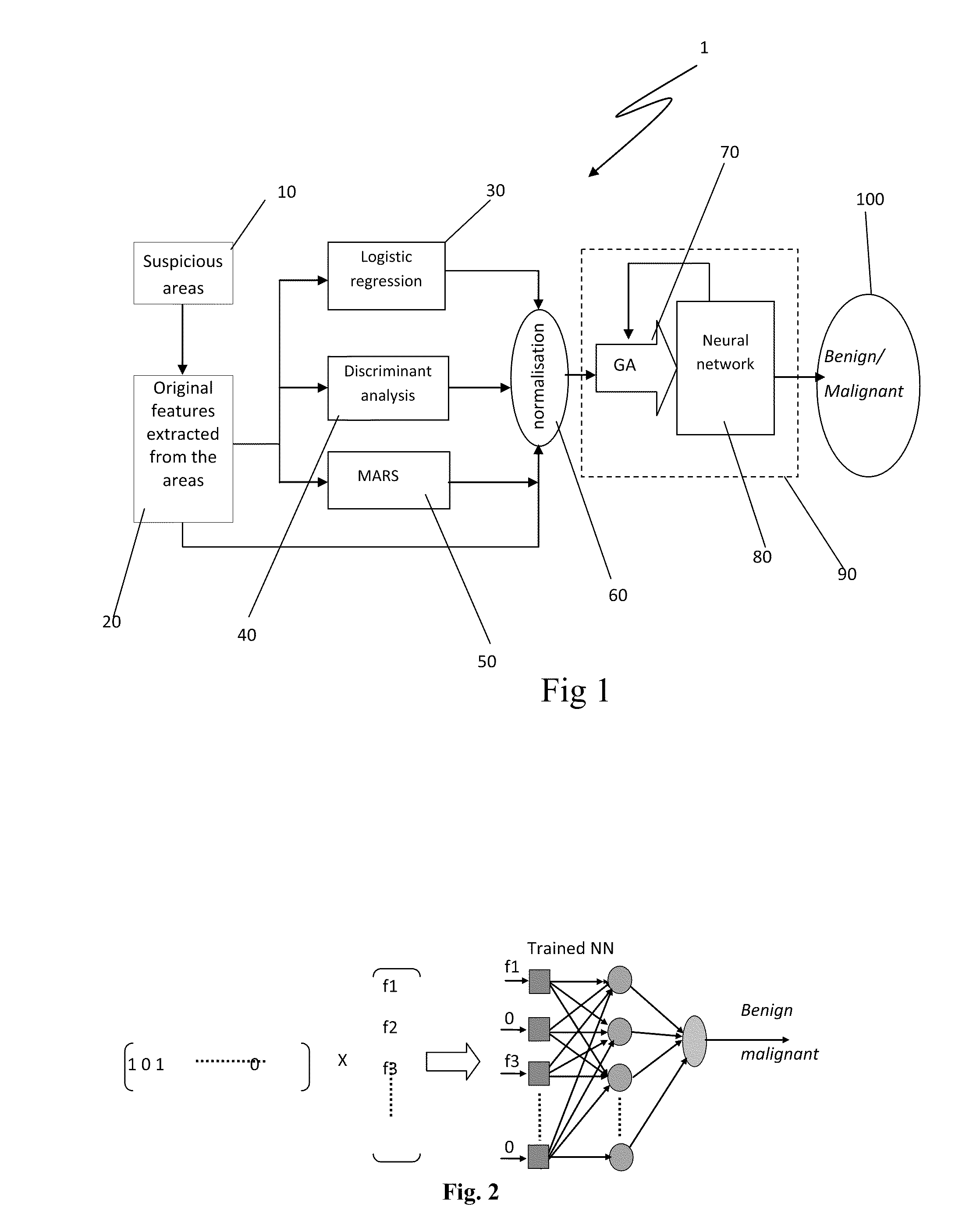
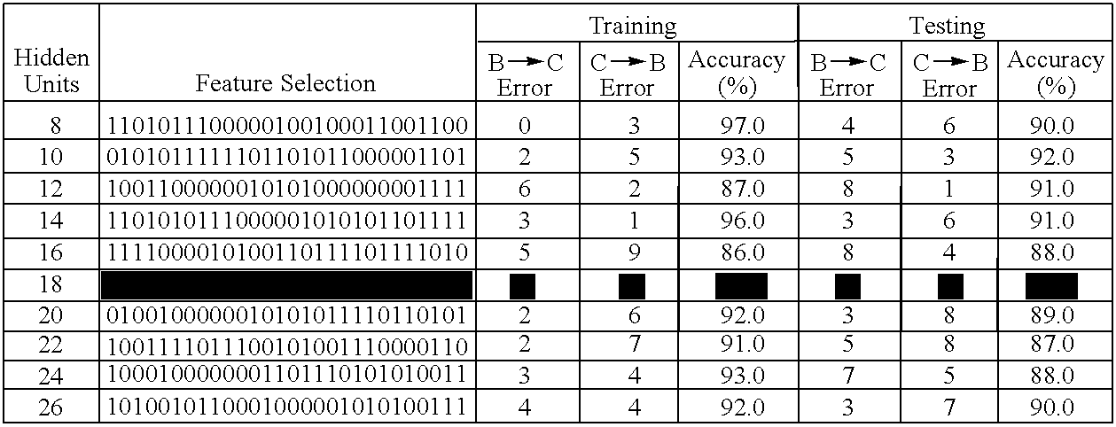
[0145]For comparing the different classification methods, the experiments were conducted using the same dataset with three different splits of 200 masses or 200 calcifications for training and testing.

[0146]In the experiment, 200 masses and 200 calcification cases were selected from the Digital Database for Screening Mammography (“DDSM”). The DDSM contains mammograms obtained from Massachusetts General Hospital, Wake Forest University School of Medicine, Sacred Heart Hospital and Washington University of St. Louis School of Medicine. The mammograms from mediolateral oblique “(MLO”) and Crabuak-Ccaudal (“CC”) views for each case have been digitized on one of four different digitizers which are DBA M2100 ImageClear, Howtek 960, Lumisys 200 Laser and Howtek MultiRad850. More information about the digitizers can be retrieved from DDSM website http: / / marathon.csee.usf.edu / Mammography / DDSM. All the cases used here had been scanned with the Howtek 960 scanner from the CC view at the full 4...


PUM


Abstract

A method and system for classifying tissue suspected of being abnormal as being malignant or benign. The method includes generating a set of selection features, performing statistical applications to generate additional selection features, generating a feature vector for the abnormal tissue, feeding the feature vector into a neural network, and obtaining a result from the neural network as to whether the abnormal tissue is malignant or benign. The method and system may be used for determining the presence of breast cancer.

Description

CROSS REFERENCE TO RELATED APPLICATIONS[0001]This application claims the benefit of U.S. Provisional Application No. 61 / 089,016, filed Aug. 14, 2008, entitled “Cancer Diagnostic Method and System,” the entire contents of which is incorporated by reference herein.FIELD OF THE INVENTION[0002]The present invention relates to improvements in methods and systems for determining whether an abnormal growth is malignant or benign.BACKGROUND OF THE INVENTION[0003]Breast cancer is the second leading cause of cancer death for all women in the world and the most common cause of death from cancer in Australian women. Early detection is the key to successful treatment and reduction of mortality. Mammography is a widely accepted way for breast cancer early detection. However, there are still 10%-30% of women who have breast cancer and undergo mammography have negative mammograms.[0004]A computer aided diagnosis (“CAD”) system can serve as the second reader to aid radiologists in breast cancer dete...

Claims


Application Information

Patent Timeline
Patent Type & Authority Applications(United States)
IPC IPC(8): G06K9/00G06V10/771
CPCG06K9/6229G06K2209/05G06K9/6292G06V2201/03G06V10/771G06F18/2111G06F18/254
Inventor ZHANG, PINGKUMAR, KULDEEP
Owner ZHANG PING
Why Eureka
Try Eureka