
External fixator for distal radius fracture

a fixator and distal radius technology, applied in the field of external fixators, can solve the problems of long time of stiffness and disability, long period of deformity, pain,

Active Publication Date: 2010-03-25
NUTEK ORTHOPAEDICS
View PDF10 Cites 2 Cited by
  • Summary
  • Abstract
  • Description
  • Claims
  • Application Information

AI Technical Summary

Benefits of technology

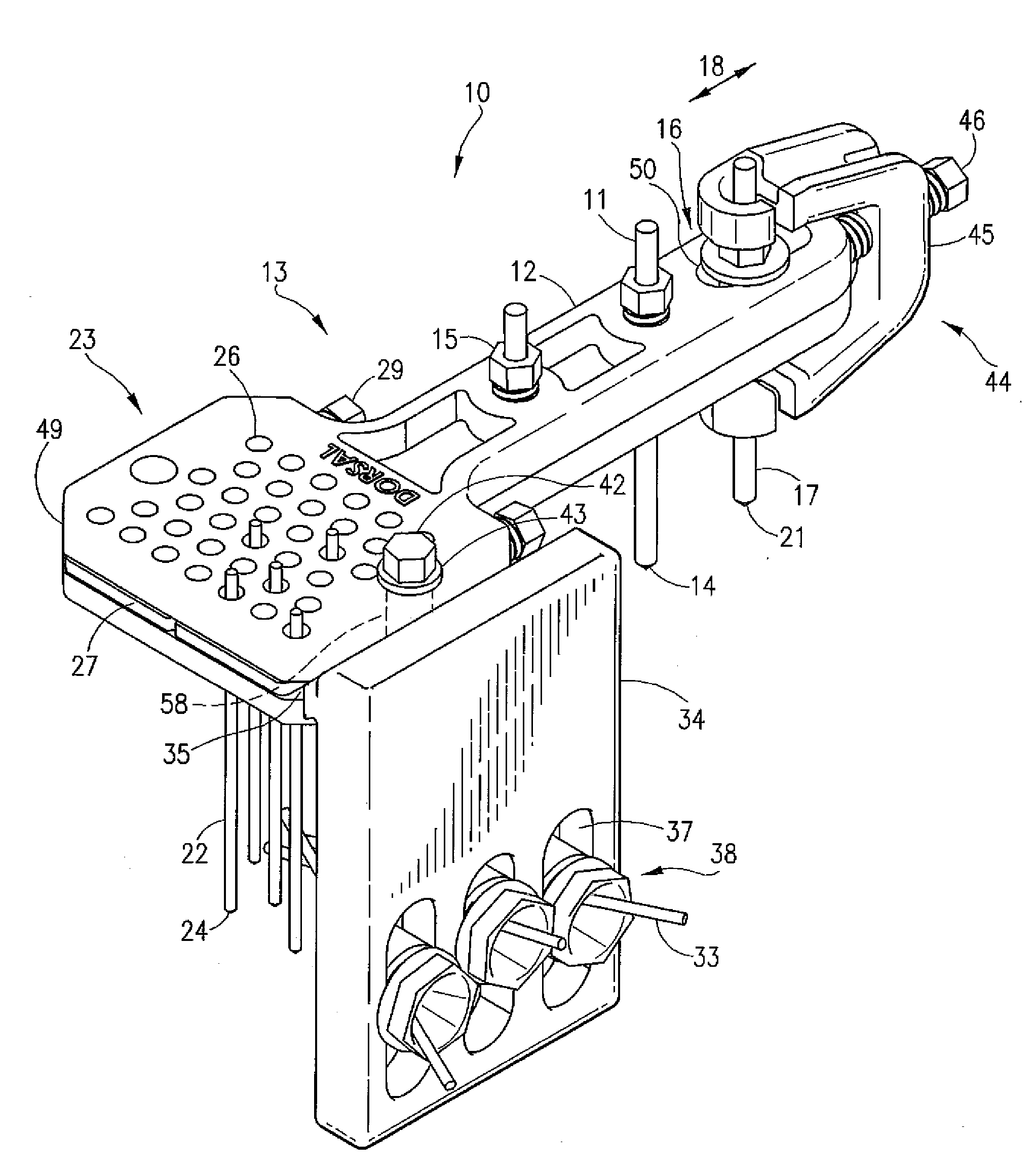
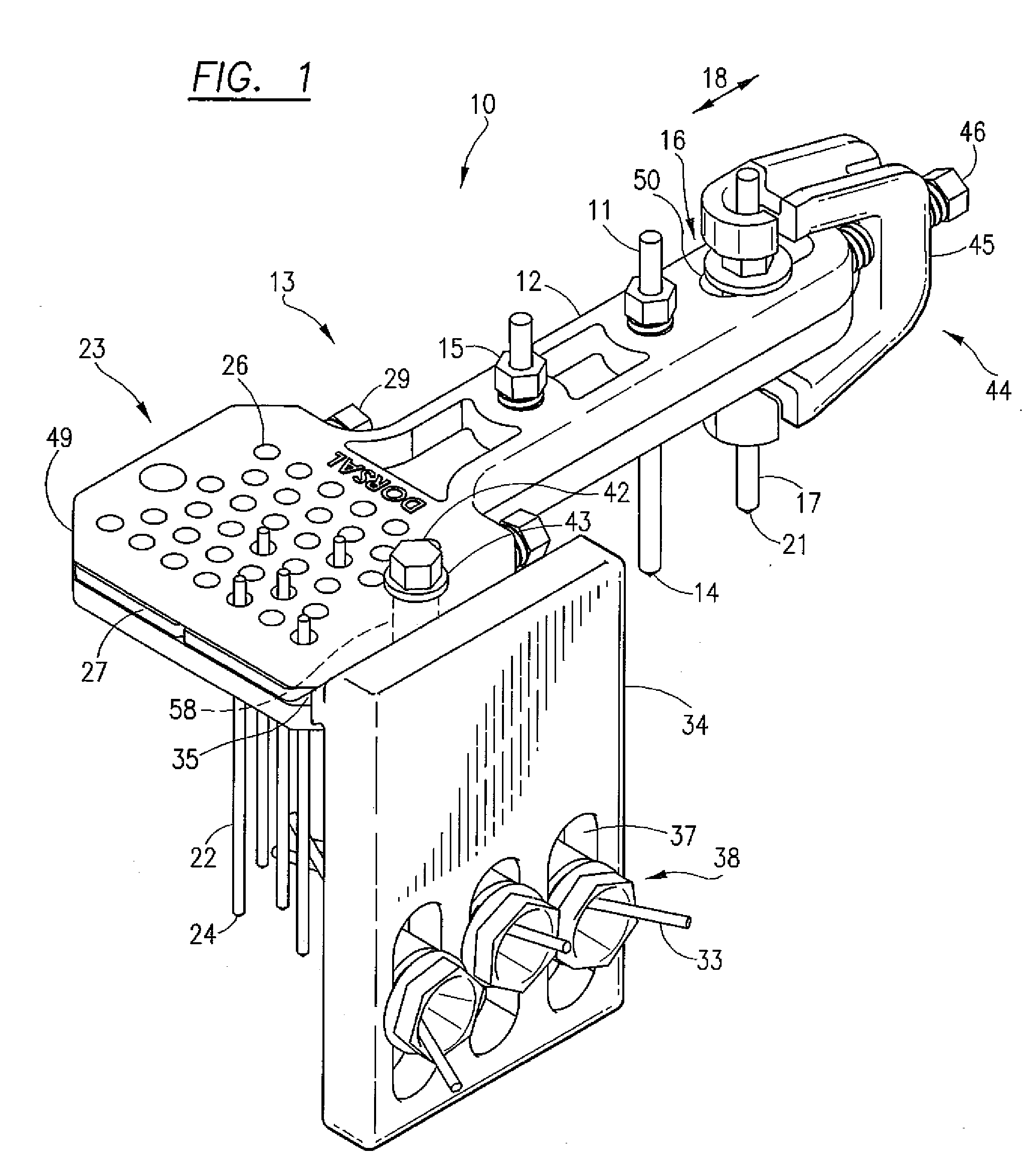
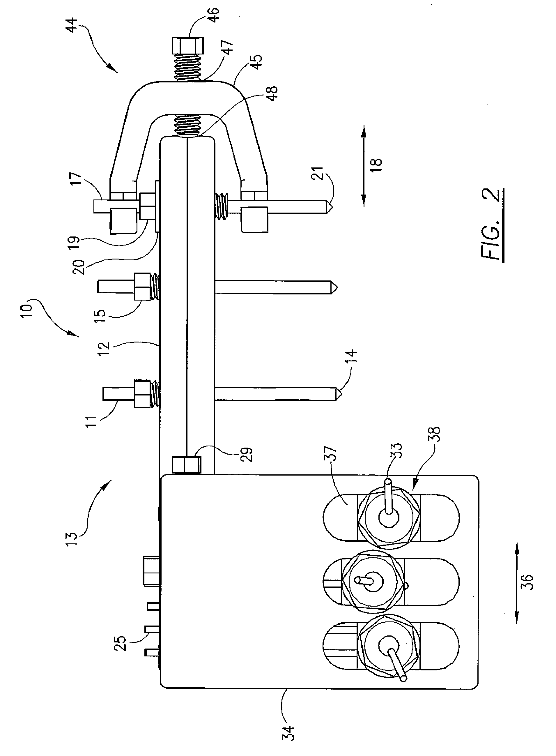
[0010]An external fixator comprising a fixator body, said fixator body having an elongated frame element at one end and a first pin mounting block at the opposite end. A second pin mounting block is orthogonally and slideably connected to one side of the fixator body. The external fixator provides a more stable structure to fix a plurality of fragments (or segments) of a fractured distal radius bone with respect to the radius bone shaft.
[0013]The second pin mounting block has one or more pin holding members, wherein each pill holding member contains a spherical pin holder (or locator) and is mounted in an elongated slot on the second pin mounting block. Each spherical pin holder allows a lateral fragment pin used to fix bone chips in place to be individually angularly disposed in a spherical angle of about thirty degrees from the perpendicular to the second pin mounting block surface and each lateral fragment pin can be screwed into a bone fragment and locked in place by tightening the pin holding member containing it. The second pin mounting block can also slide laterally (backwards and forwards in the same direction as the radius bone) for additional positioning of the horizontal fragment pins.
[0016]Using the present invention as an external fixator for a radius fracture allows for immediate post operative wrist and finger function and motion from dorsal to volar function, radial to ulnar deviation, supination, and pronation.

Problems solved by technology

A problem with this method is that it sometimes results in inadequate internal fixation, which can cause deformity, pain, and prolonged disability.
While this type of fixation often provides an improvement over conventional casting techniques in the management of severe fractures of the distal radius, immobilization of the radiocarpal joint during the treatment period typically results in a long period of stiffness and disability after the external fixation device is removed.

Method used


Image

  • External fixator for distal radius fracture
  • External fixator for distal radius fracture
  • External fixator for distal radius fracture

Examples


Embodiment Construction

[0023]An external fixator 10, built in accordance with the present invention, will now be described, with an initial reference to FIG. 1, FIG. 2, and FIG. 3. The external fixator 10 is configured for surgical attachment to the shank portion of a radius bone (not shown) by means of two mounting pins 11, extending downward from an elongated frame element 12 of a fixator body 13, with the pointed end 14 of each mounting pin 11 being screwed into the bone. In the elongated frame element 12, a pin clamping screw 15 is used to hold each mounting pin 11 in a fixed relationship with the fixator body 13. Each pin clamping screw 15 extends within an elongated frame element aperture in the elongated section and includes a number of flexible sections that move inward engaging the pin 11 extending through the pin clamping screw 15 as that pin clamping screw 15 is driven into engagement with said elongated frame element aperture. Near the proximal end 16 of the elongated frame element 12, a slidi...


PUM


Abstract

An external fixator for a fractured distal radius including a first plurality of vertical passages for pins extending downward from a first pin mounting block into one or more bone fragments and one or more frame element apertures extending from an elongated frame element for pins extending downward from the device into the shaft of the radius. In addition, the elongated frame element includes a sliding pin slot with a sliding structure allowing a single pin to be moved to provide extension between the fragments and the shaft. A sliding internal plate including holes aligned with the first plurality of vertical passages is moved by a pair of setscrews to clamp the pins extending through the first plurality of vertical passages. The device also includes a second pin mounting block that extends downward parallel to the pins extending from first pin mounting block to hold pins directed laterally into the fragment(s).

Description

BACKGROUND OF THE INVENTION[0001]1. Field of the Invention[0002]This invention relates to devices for external fixation of fractured bones, and, more particularly, to an external fixator in which bone setting pins are clamped from a plurality of angles for fixation of multiple fragments of an end portion of a radius bone.[0003]2. Description of the Related Art[0004]The fracture of the distal radius is one of the most common human fractures, occurring in as many as 350,000 people per year in the United States alone. The conventional processes both for reducing such a fracture and for maintaining the bones in proper alignment during the subsequent healing process involves applying and maintaining an extension force across the fracture, with ligamental taxis being relied upon to hold the bones in place. The process for treating a fractured distal radius is described in the 1901 edition of Gray's Anatomy in the following manner, “The treatment consists of flexing the forearm, and making...

Claims


Application Information

Patent Timeline
Patent Type & Authority Applications(United States)
IPC IPC(8): A61B17/00
CPCA61B17/66
Inventor HAJIANPOUR, MOHAMMED A.
Owner NUTEK ORTHOPAEDICS
Learn More