
C-Type Lectin Fold as a Scaffold for Massive Sequence Variation

a lectin and sequence variation technology, applied in the field of binding proteins, can solve the problems of limited variability in the sequence of proteins, immunoglobulins, and amino acid residues with side chains that are not exposed to the exterior solvent, and achieve the effects of stable structure, high tolerance, and stable structur

Inactive Publication Date: 2010-03-18
RGT UNIV OF CALIFORNIA
View PDF2 Cites 7 Cited by
  • Summary
  • Abstract
  • Description
  • Claims
  • Application Information

AI Technical Summary

Benefits of technology

[0021]The availability of a scaffold to present diverse binding sites permits the generation of binding proteins with different specificities and affinities for binding a wide number of different target molecules, particularly biomolecules. The binding proteins may be used to bind, and thus detect, identify, localize or modify, such target molecules.
[0079]A modified scaffold or binding protein attached to a toxin, or pro-drug form thereof, may be used to decrease the viability of, or to kill, cells which express a cell surface molecule bound by the modified scaffold or protein. Preferably, the cells are cancer cells, such as those of a mammal, preferably a human.

Problems solved by technology

But variability in the sequence of a protein, like an immunoglobulin, is often limited by the effects of variability on protein folding and the resulting final 3D shape.
Amino acid residues with side chains that are not exposed to the exterior solvent are often limited in variability because as part of the protein's interior they must “fit” within the interior space as dictated by other amino acid residues.
The usefulness of immunoglobulins as manipulable binding proteins is limited, however, by the nature of the immunoglobulin framework, which requires two polypeptides to form the complete ligand- or antigen-binding site.
This results in a number of disadvantages: the need to manipulate rather large polypeptides, the need for complicated molecular cloning to diversify a binding site; and the complication of modifying six different CDRs.

Method used


Image

  • C-Type Lectin Fold as a Scaffold for Massive Sequence Variation
  • C-Type Lectin Fold as a Scaffold for Massive Sequence Variation
  • C-Type Lectin Fold as a Scaffold for Massive Sequence Variation

Examples


example 1

Crystal Structures of MTD Variants

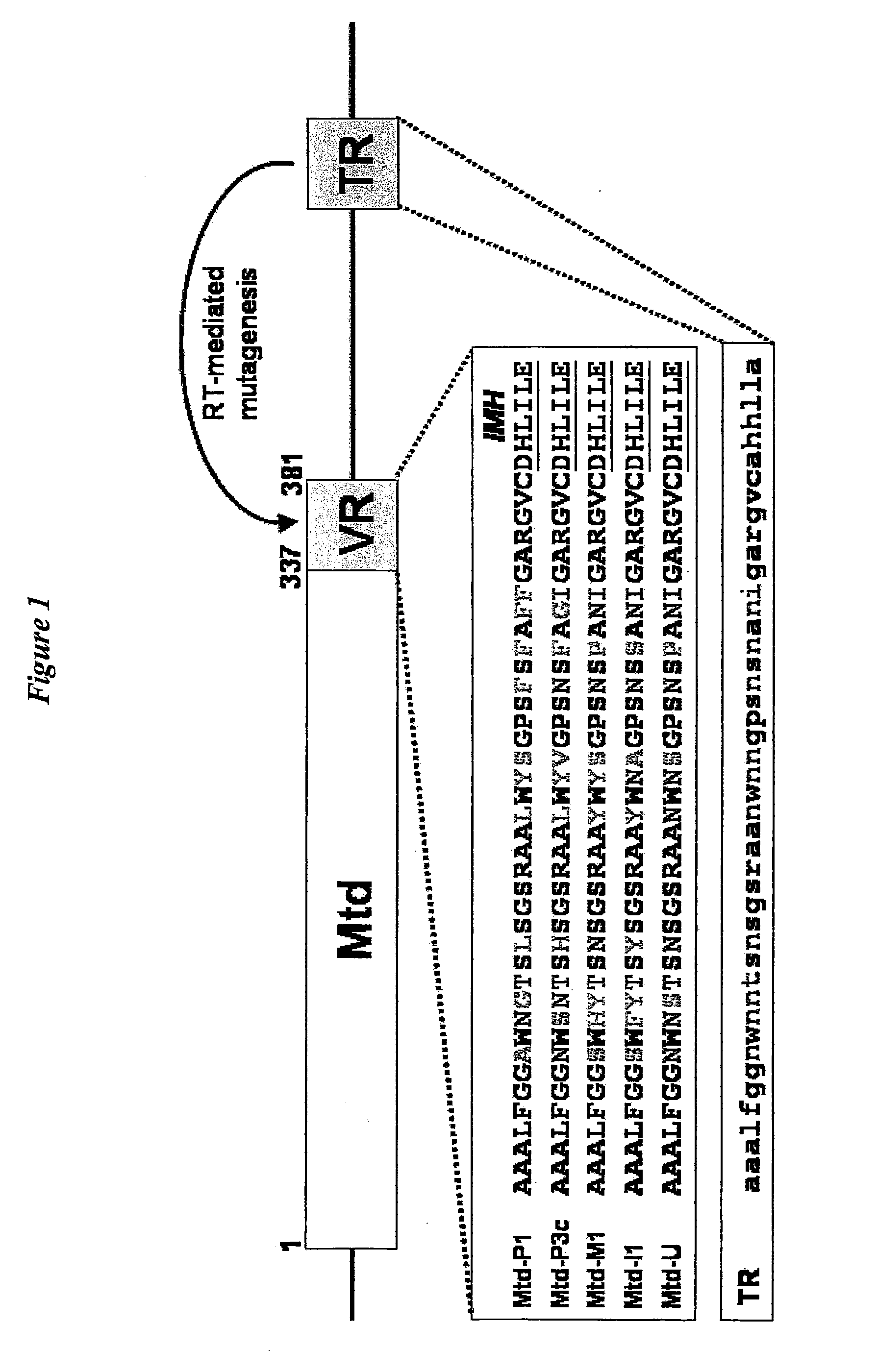
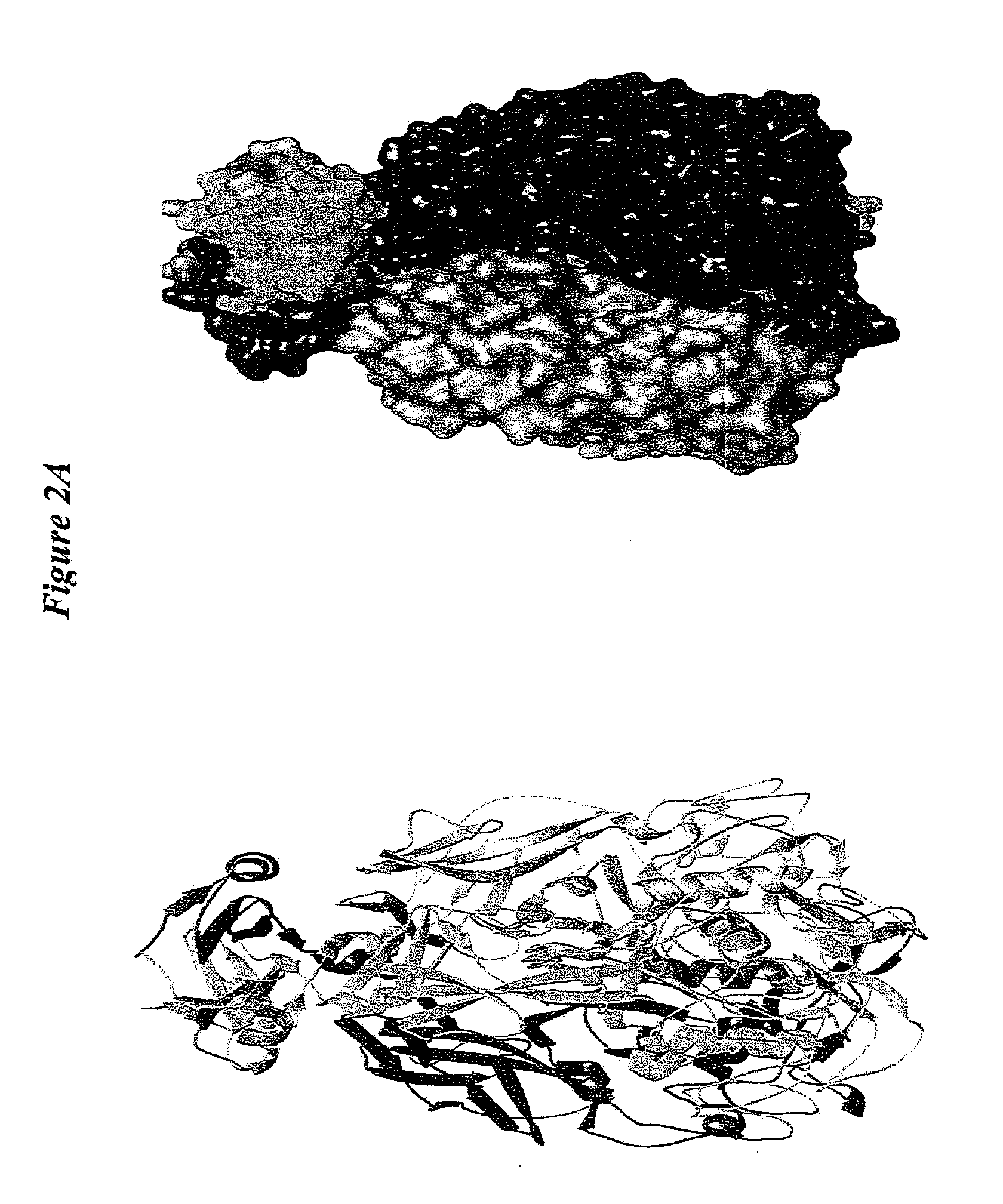
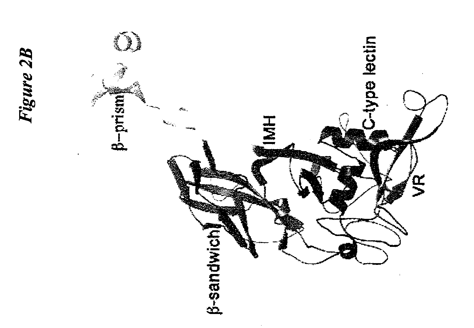
[0147]Structural comparison of Mtd-P1,-3c, -M1, I1, and -N1 were used to discover that the main chain conformation of the CTL domain is remarkably invariant, despite half of the variable residues being on loop regions (FIG. 3C). The binding site in these variants is highly well ordered, having average main chain B-factors ranging from ˜9 Å in Mtd-P1 to −24 Å2 in Mtd-M1 and with density visible for all but one side chain (Phe-346 in Mtd-I1). Providing stabilization to these loops in Mtd are two features unique to the Mtd CTL-fold, namely the two inserts and trimeric assembly.

[0148]The inserts form hydrogen bonds to VR, including three to side chains of three invariant serines in VR. Ser-270 and Glu-267 from the second insert form hydrogen bonds to the invariant VR residues Ser-351 and Ser-353, respectively (FIG. 3D), and main chain atoms of the first insert form hydrogen bonds to invariant VR residue Ser-365 (not depicted). These interactions are sup...

example 2

Basis of MTD to Ligand Interactions

[0150]To understand the basis of Mtd interactions with its ligand, a cell surface receptor, we characterized association between Mtd-P 1 and the Bordetella receptor pertactin. The pertactin ectodomain (Prn-E) was incubated with Mtd variants and found by a coprecipitation assay to associate most strongly with Mtd-P1 but also with Mtd-3c and Mtd-M1. As a measure of specificity, Prn-E was not found to associate with Mtd-I1 or Mtd-N1. The three Mtd variants that are found to bind pertactin have in common the variable residue Tyr-359, previously shown by sequence comparison to be a consistent determinant for pertactin interaction. The presence of a tyrosine residue in the binding pocket is consistent with the presence of a number of hydrophobic surface-exposed patches on Prn-E (see Emsley, P., et al. Structure of Bordetella pertussis virulence factor P.69 pertactin. Nature 381, 90-2 (1996)). The maintenance of Pm affinity in some of these Mtd variants a...

example 3

CTL-Fold in Other DGRs

[0152]A number of other putative DGRs have been identified in phage and bacterial genomes. These resemble the Bordetella phage DGR in having sequence-related reverse transcriptases, similar arrangements of VR and TR, adenines constituting the main differences between VR and TR, and IMH-like elements at the end of VR. However, the putative variable proteins have no obvious sequence relationship to Mtd or other proteins. Because there appears to be no genetic requirement for VR and its IMH element to be positioned at the very C-terminus of a protein, the variations in positioning likely reflects the necessities of protein binding requirements as specified by the CTL-fold. Despite the low sequence identity among these proteins (˜17%), we have been able to use the structure of Mtd along with considerations about variability to construct a sequence alignment consisting of the β2β3β4β4′ sheet of the CTL-fold (see FIG. 5). Most notably, the invariant Mtd binding site ...


PUM

PropertyMeasurementUnit
lengthaaaaaaaaaa
nucleic acidaaaaaaaaaa
surfaceaaaaaaaaaa

Abstract

This invention provides a class of binding proteins with a range of binding specificities and affinities based upon variation at select amino acid positions within a scaffold. The variable positions may be readily modified to produce a library of binding proteins with different binding specificities and affinities. The library may be screened to identify one or more as binding a ligand of interest. Compositions comprising the binding proteins, as well as methods of using the binding proteins are also provided.

Description

CROSS REFERENCE TO RELATED APPLICATIONS[0001]This application is a continuation application of U.S. application Ser. No. 11 / 027,323 filed Dec. 31, 2004, now pending. The disclosure of the prior application is considered part of and is incorporated by reference in the disclosure of this application.GRANT INFORMATION[0002]This invention was made with government support under Grant Nos. T32 GM008326, F31 AI061840 and F32 AI49695 awarded by the National Institutes of Health. The government has certain rights in the invention.BACKGROUND OF THE INVENTION[0003]1. Field of the Invention[0004]This invention relates to a class of binding proteins with a range of binding specificities and affinities based upon variation at select amino acid positions within a scaffold. The variable positions may be readily modified to produce a variety of binding proteins with different binding specificities and affinities. This range of proteins may be screened to identify one or more as binding a target mole...

Claims


Application Information

Patent Timeline
Patent Type & Authority Applications(United States)
IPC IPC(8): A61K38/16C07K14/42C07H21/04C12P21/00G01N33/53A61P35/00
CPCC07K14/195C07K14/001A61P35/00
Inventor GHOSH, PARTHOMCMAHON, STEPHENMILLER, JASONLAWTON, JEFFREY
Owner RGT UNIV OF CALIFORNIA
Learn More