
Rapid magnetic flow assays

a magnetic flow and assay technology, applied in the field of molecular biology and medical science, can solve the problems of easy breakage of molecular tethers, achieve the effect of full antigenicity and binding integrity, rapid methods for forming target specific detection complexes, and wide range of effects

Inactive Publication Date: 2009-06-11
PERKINELMER HEALTH SCIENCES INC
View PDF99 Cites 71 Cited by
  • Summary
  • Abstract
  • Description
  • Claims
  • Application Information

AI Technical Summary

Benefits of technology

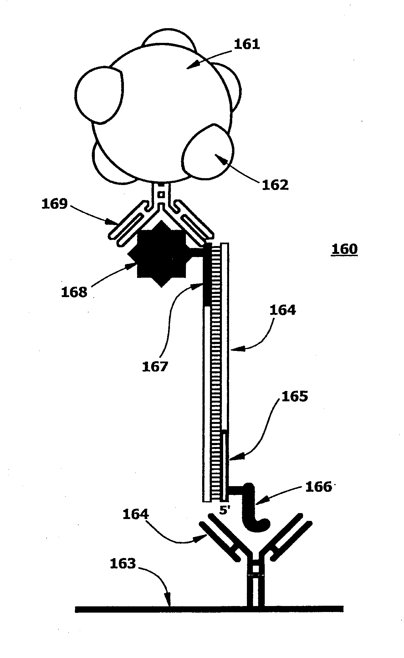
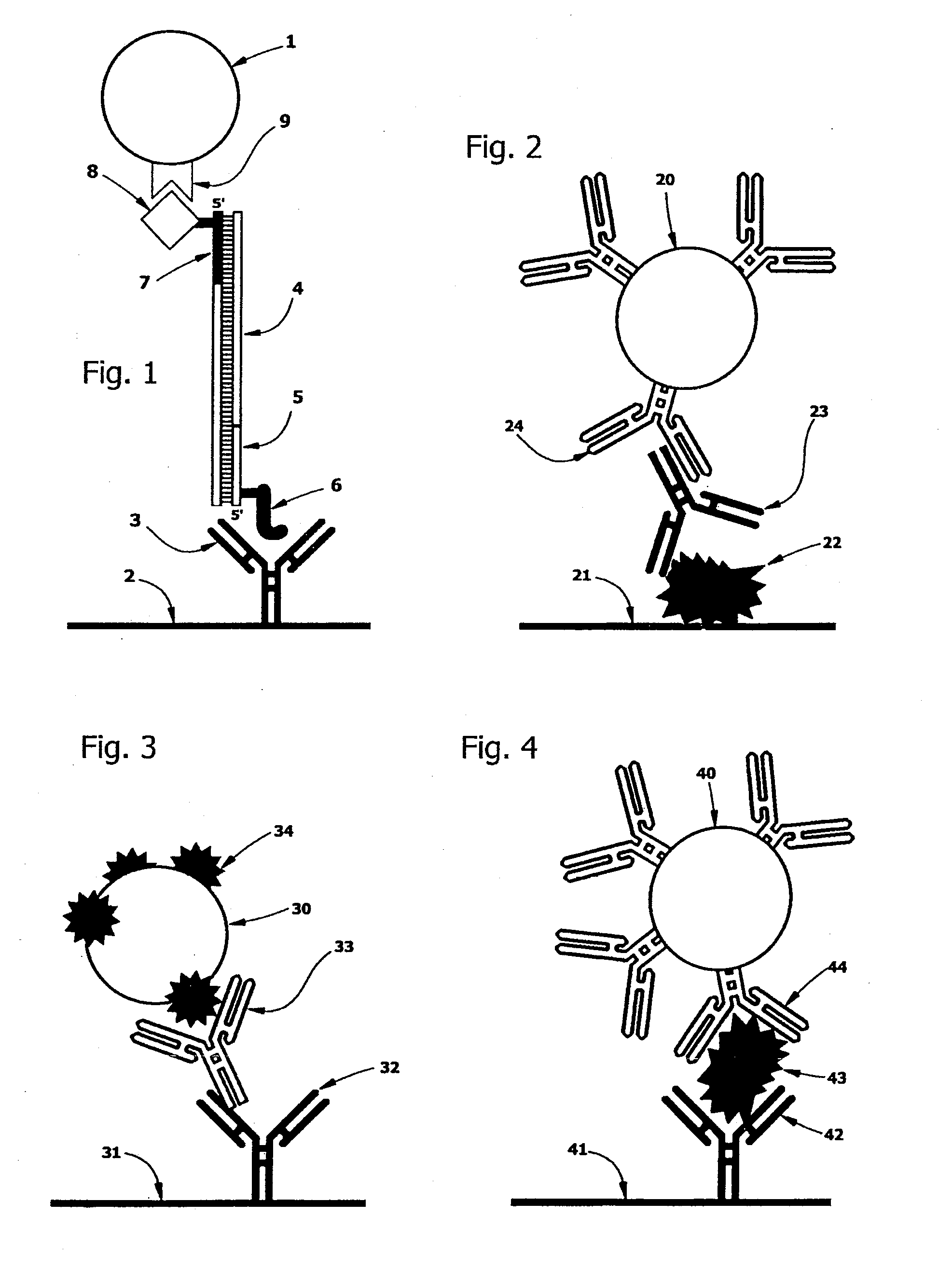
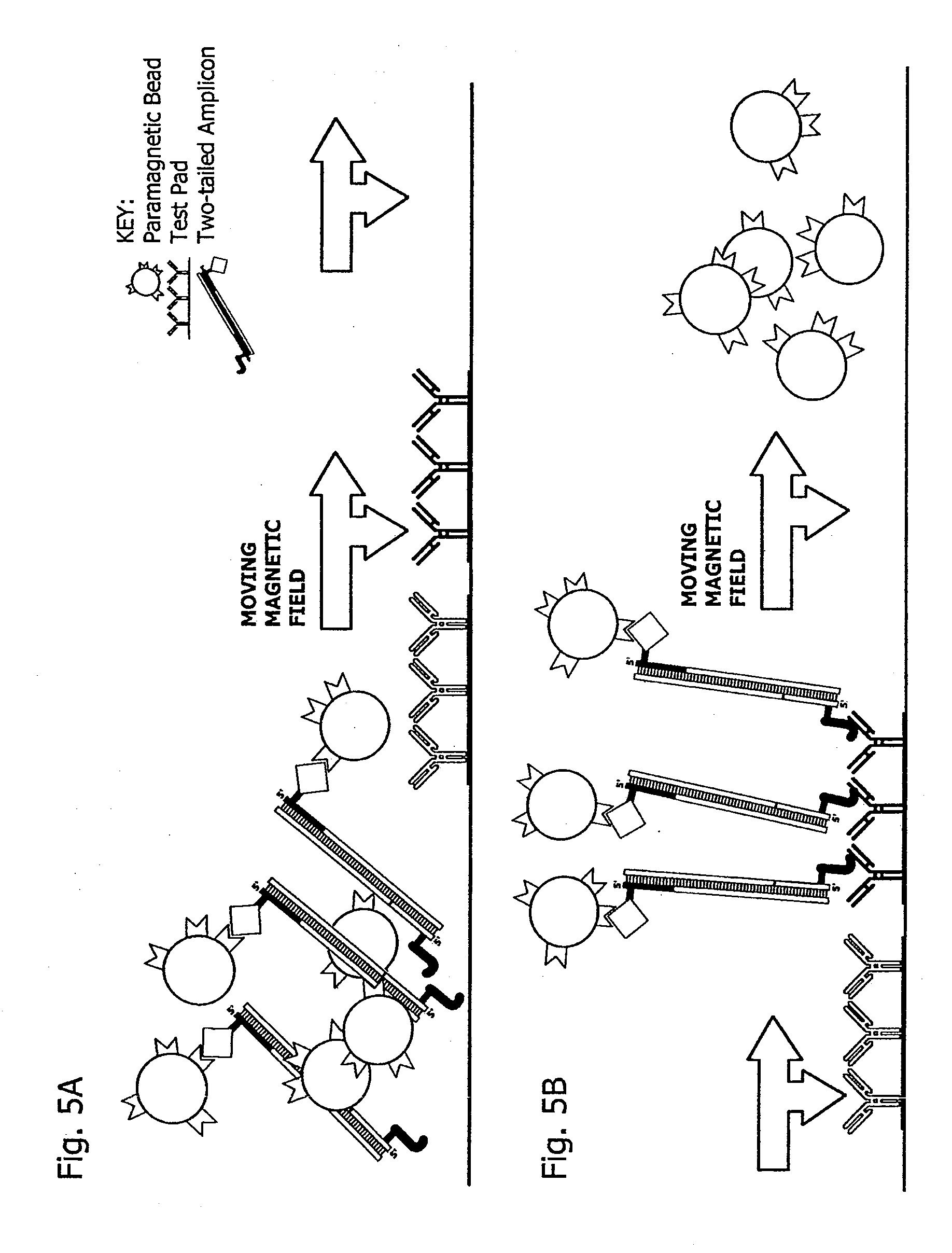
[0021]Surprisingly, ligand-tagged paramagnetic microbeads are readily extracted from a moving magnetic field by formation of molecular tethers with solid phase substrates coated with affinity ligand-binding molecules.

Problems solved by technology

At odds with this finding, the prior art has taught that such molecular tethers are easily broken and that stationary magnetic fields are needed to keep paramagnetic beads immobilized during washing and separation of bound and unbound beads.

Method used


Image

  • Rapid magnetic flow assays
  • Rapid magnetic flow assays
  • Rapid magnetic flow assays

Examples


example 1

A) Preparation of Primer Sets

[0189]Reverse primers were first prepared and HPLC purified. Peptides were derivatized with n-terminal hydrazine before use. Oligonucleotides were treated with succinimidyl 4-formylbenzoate in formamide and then reacted with the hydrazine derivatized peptides to form hapten-tagged primers.

[0190]The following peptidyl hapten-tagged primers were used.

5′-Peptidyl OligomersPri-merPrimer Sequence*Peptide Sequence**ACGCCAGTACGATATTCAG(HNA) EQKLISEEDL(SEQ ID NO:1)(NH2) (SEQ ID NO:8)BACCTGGACATCACGGCTTTCAAC(HNA) YPYDVPDYA(SEQ ID NO:2)(NH2) (SEQ ID NO:9)CCCTATTGCAGAGCGAATGAC(HNA) YTDIEMNRLGK(SEQ ID NO:3)(NH2) (SEQ ID NO:10)DTGAACTCCATTAACGCCAGA(HNA) CEEEEYMPME(SEQ ID NO:4)(NH2) (SEQ ID NO:11)ECGACCTGACCAAATGCCAG(HNA) TDFYLK (NH2)(SEQ ID NO:5)(SEQ ID NO:12)FCCTATAACAGCACCCACTATACGG(HNA) DTYRYI (NH2)(SEQ ID NO:6)(SEQ ID NO:13)GCTCTGCGAGCATGGTCTGG(HNA) QPELAPEDPED(SEQ ID NO:7)(NH2) (SEQ ID NO:14)

[0191]These peptide epitopes were selected based on the availability of...

example 2

[0198]PCR amplification was performed in a microfluidic device as follows:

[0199]A microfluidic device was built from stencil-cut laminates. Before final assembly, biotin- and hapten-tagged primer pairs, dATP, dCTP, dGTP and dTTP, TAQ polymerase, and a matrix consisting of TritonX100, BSA, PEG and Trehalose plus magnesium chloride were deposited in the amplification channel or chamber and dried in place under vacuum. Streptavidin-coated magnetic beads (Dynal MyOne Streptavidin Cl, Carlsbad Calif.) were spotted and dried in a chamber adjoining the amplification channels or chambers. Test pad areas in the detection chamber were stenciled (see FIG. 21 for general approach) and gas plasma treated, before antibody solutions were applied and dried in place. Antibody spots were blocked with StabilCoat (SurModics, Eden Prairie Minn.). The device was then treated with a TritonX100:BSA buffer to passivate untreated plastic surfaces.

The following reagents were also prepared:

Lysis Buffer

[0200]4....

example 3

[0222]A result of an assay in which the targets of Example 2 were extracted, amplified and detected is shown in FIG. 6.


PUM

PropertyMeasurementUnit
Areaaaaaaaaaaa
Affinityaaaaaaaaaa
Paramagnetismaaaaaaaaaa

Abstract

Disclosed is an improvement in methods for nucleic acid and immunological bioassays. The methods comprise a step for “sweeping” paramagnetic bead: target molecule complexes so as to capture them with an affinity capture agent on a test pad by moving a magnetic force field from outside to inside the test pad area so as to bring into contact the paramagnetic complexes with the capture agent, while sweeping any unbound paramagnetic material off the test pad by moving the magnetic field from inside to outside the test pad area. Surprisingly, the paramagnetic complexes are rapidly affinity-extracted from the moving magnetic field.

Description

CROSS-REFERENCE TO RELATED APPLICATIONS[0001]This application is a continuation of International PCT Patent Application No. PCT / US2007 / 006585, filed Mar. 15, 2007, now pending, which claims the benefit under 35 U.S.C. 119(e) of U.S. Provisional Patent Application No. 60 / 782,649, filed Mar. 15, 2006, and U.S. Provisional Patent Application No. 60 / 844,811, filed Sep. 14, 2006. These applications are incorporated herein by reference in their entireties.STATEMENT OF GOVERNMENT INTEREST[0002]This invention was made with government support under Contract No. UO1 AI061187, awarded by the National Institutes of Health. The government has certain rights in this invention.STATEMENT REGARDING SEQUENCE LISTING[0003]The Sequence Listing associated with this application is provided in text format in lieu of a paper copy, and is hereby incorporated by reference into the specification. The name of the text file containing the Sequence Listing is 660115—455C1_SEQUENCE_LISTING.txt. The text file is 6...

Claims


Application Information

Patent Timeline
IPC IPC(8): C12Q1/68C07H21/04B01J19/00
CPCB01F11/0071B01F13/0059C12Q1/686C12Q1/6834B01L2400/0655B01L2400/0638B01L2400/0481B01L3/5027B01L3/502723B01L3/50273B01L3/502738B01L3/502746B01L7/52B01L2200/082B01L2200/10B01L2200/16B01L2300/0663B01L2300/0816B01L2300/0867B01L2300/087B01L2300/0887C12Q2563/131C12Q2525/197C12Q2563/143C12Q2565/629B01F31/65B01F33/30
Inventor KOKORIS, MARKNABAVI, MELUDBREIDFORD, WAYNE L.GERDES, JOHNMORDUE, STEPHENBATTRELL, C. FREDERICK
Owner PERKINELMER HEALTH SCIENCES INC
Learn More