
Method for mapping image reference points to facilitate biopsy using magnetic resonance imaging

a magnetic resonance imaging and reference point technology, applied in the field of magnetic resonance imaging, can solve the problems of unfavorable tissue biopsy, unfavorable tissue biopsy, and difficult to determine the tissue targeted for biopsy, so as to improve the image quality and quickly assess the accuracy of targeting

Inactive Publication Date: 2009-06-04
AURORA HEALTHCARE US CORP
View PDF14 Cites 24 Cited by
  • Summary
  • Abstract
  • Description
  • Claims
  • Application Information

AI Technical Summary

Benefits of technology

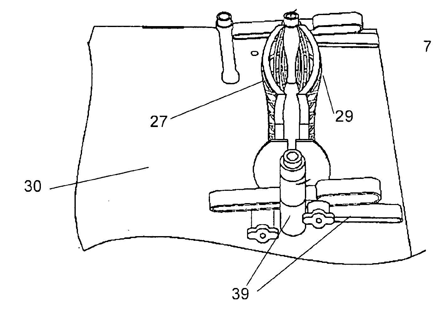
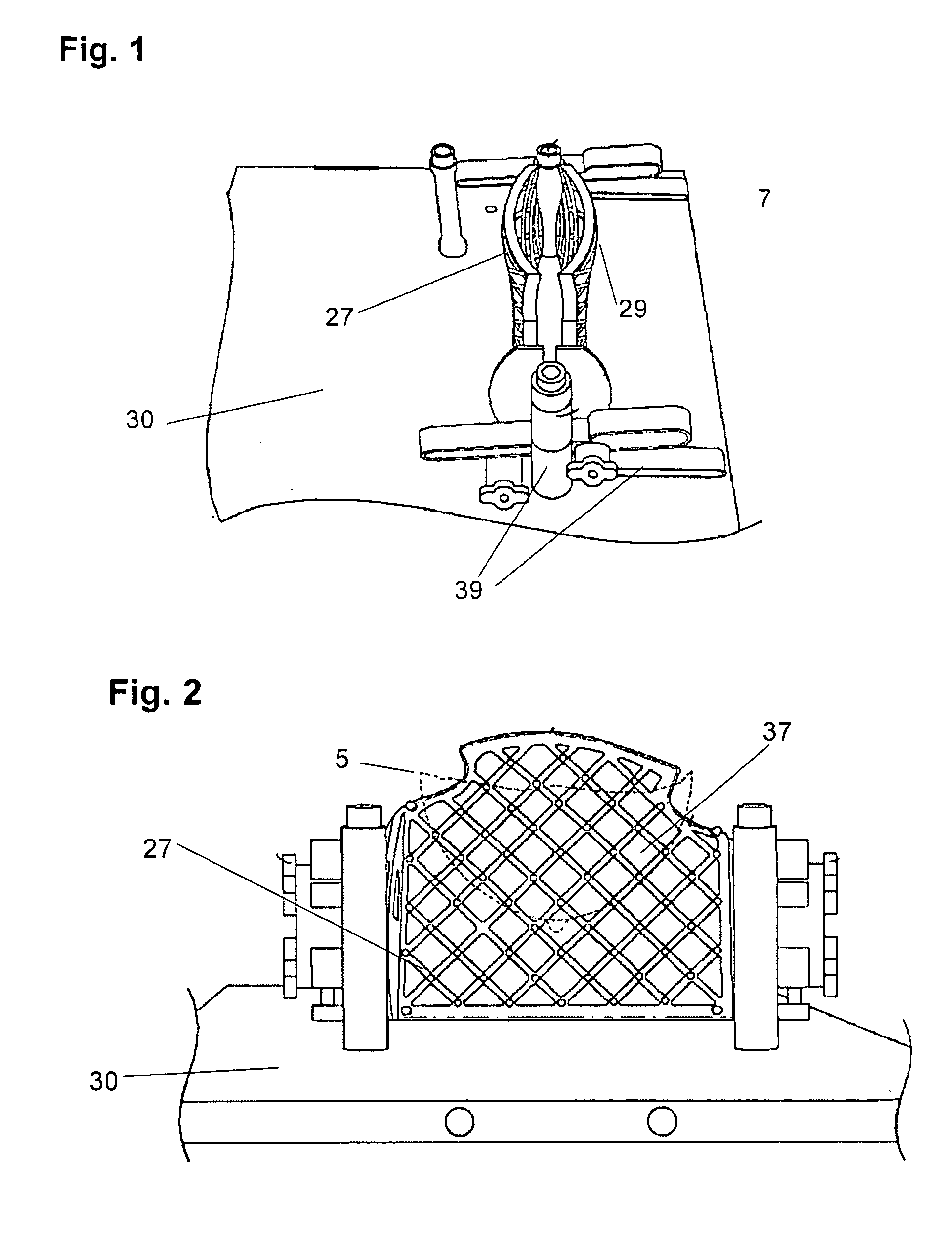
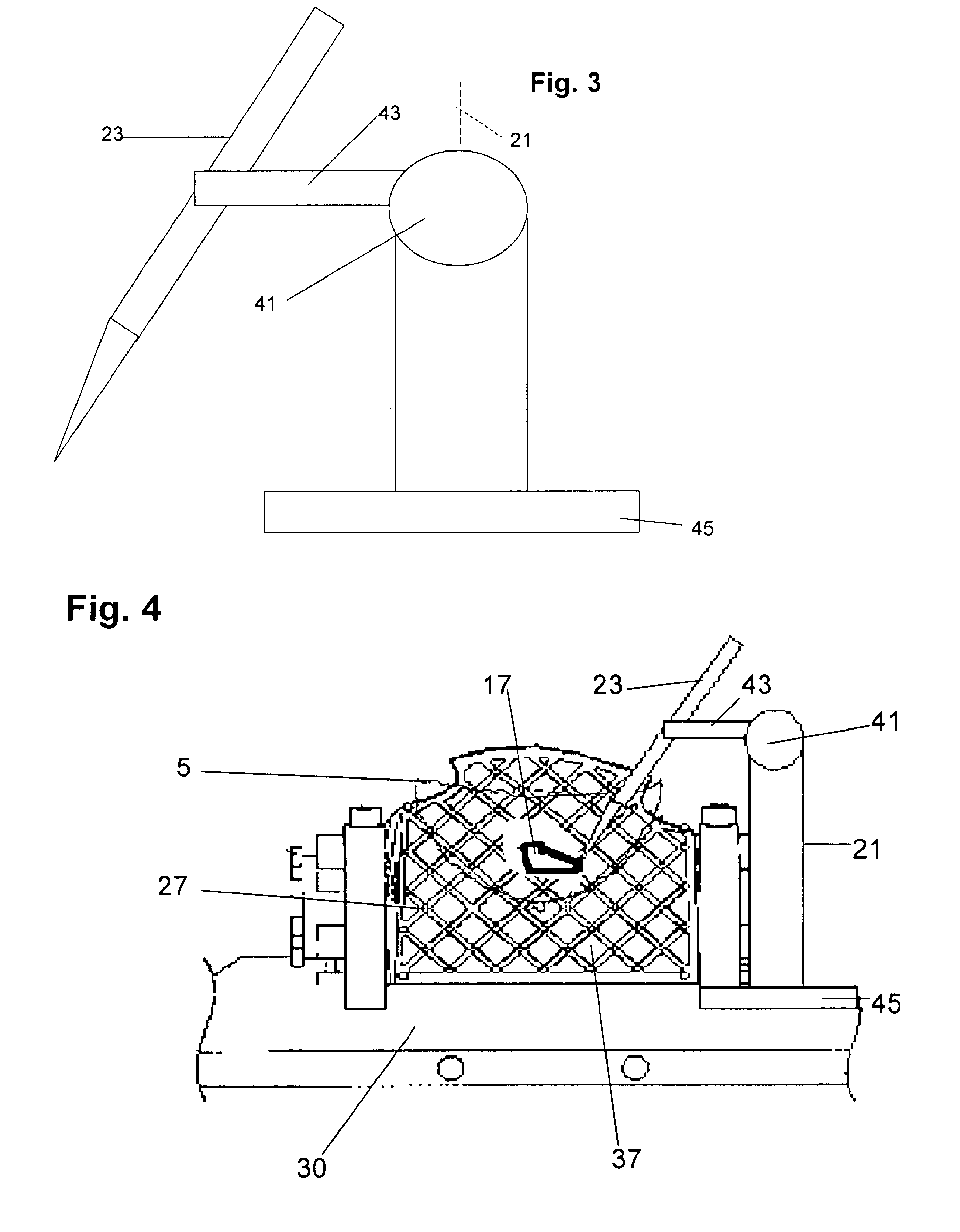
[0021]In order to provide a more versatile and yet effective way to ensure that a biopsy needle holder (or a similar device for deploying a modality) can be accurately positioned relative to tissue that may be identified in an earlier image but currently not visible, the present disclosure proposes that the needle holder or a fiducial marker on the needle holder form a point of reference for positioning, aiming and deploying the modality so as to accurately intersect the target tissue. The needle holder need not be positioned relative to the patient table, or fixed in space, and instead can be moved. The needle holder is provided with a displacement measurement encoder, or alternatively is arranged such that its displacement in position from one image to another can be measured. Then provided that the breast remains in position, the change in position of the needle holder is accommodated. A new trajectory is calculated based on the new position of the needle holder, which is used as a movable reference, and the holder is aimed to deploy the modality to intersect the target tissue accurately.

Problems solved by technology

The foregoing technique of fiducial marking and planning is theoretically effective but is unwieldy.
The technique also can be unsure if the tissue targeted for biopsy is not readily visible in follow-up image sequences.
This problem is likely to occur when attempting to biopsy a lesion that was only made to appear using a contrast agent to render the lesion visible in an image.
After perfusion with a contrast agent, which may reveal a lesion or similar tissue structure of interest, washout of the contrast agent causes the contrast at the tissue structure to be lost and the detail used to identify the tissue structure is lost.
It might be possible to repeatedly perfuse the patient, but this is not recommended.

Method used


Image

  • Method for mapping image reference points to facilitate biopsy using magnetic resonance imaging
  • Method for mapping image reference points to facilitate biopsy using magnetic resonance imaging
  • Method for mapping image reference points to facilitate biopsy using magnetic resonance imaging

Examples


Embodiment Construction

[0056]FIG. 8 shows the main elements of an exemplary embodiment of the invention, wherein a patient 1 is placed in a prone position on a supporting table 3 or other suitable support. The patient is supported at the shoulders and torso; however a gap or opening in the support permits the breasts 5 to depend downwardly, presented for imaging. The supporting table is arranged so that the patient is held stationary relative to the table. The table can be translated into and out of the coils of a NMR / MRI imaging apparatus 33, show as a cabinet in FIG. 8. In the lumen of the coils (not shown in FIG. 8), the breasts are imaged. When the table is retracted to move the patient back to the position shown in FIG. 8, the patient is accessible for various procedures, including imaging-related activities to assist in procedures and to exploit location sensitive modalities such as the injection of a contrast agent, diagnostic procedures such as biopsy, therapeutic targeted application of nuclear o...


PUM


Abstract

In NMR / MRI imaging, a location is noted for a point in the imaged space, and referred to a reference location so that the point in imaged space is known thereafter, without the need to locate the point again in further imaging steps. For breast cancer diagnosis and biopsy, a breast holding fixture immobilizes the breast. A volumetric image is taken encompassing a portion of the breast. In the same or a subsequent image, a fiducial mark is detected to determine the position of a holder for a biopsy tool or other modality. The tissue feature can be a tumor, cyst or tubal lesion, made temporarily visible in the image by perfusion with a contrast agent. After the contrast agent dissipates, the location of the tissue feature can still be determined by reference to the position of the fiducial marker, which is optional adjustable by post-imaging metered displacement.

Description

BACKGROUND OF THE INVENTION[0001]1. Field of the Invention[0002]The invention relates to magnetic resonance imaging (MRI), particularly for imaging and mapping tissue structures within the breast, and more particularly to facilitating accurate collection of biopsy tissue samples.[0003]2. Related Art[0004]Nuclear magnetic resonance imaging (NMR or MRI) is a well developed technique for providing images that can be encoded and displayed as image planes or slices through a volume of tissue, or as two dimensional views or projections of the volume. The data is collected by distinguishing the electromagnetic responses of a nuclei from different tissues, and their respective spatial positions in the imaged volume. The variations in local responses are mapped as corresponding variations in displayed luminance, color or other characteristics, applied to displays that are generated by the imaging system to represent an image of the internal tissue structures.[0005]MRI is useful for visualizi...

Claims


Application Information

Patent Timeline
IPC IPC(8): A61B5/05
CPCA61B5/055A61B10/0233A61B19/201A61B2019/5236A61B2019/205A61B2019/507A61B19/50A61B34/10A61B90/11A61B90/17A61B2034/107A61B2090/374
Inventor HARMS, STEVEN E.JIANG, AIPINGHONG, XIAOLE
Owner AURORA HEALTHCARE US CORP
Learn More