Eureka AIR delivers breakthrough ideas for toughest innovation challenges, trusted by R&D personnel around the world.

Surgery accessory and method of use

a technology of surgical accessories and methods, applied in the field of remote viewing surgery, can solve the problems of not being accepted, the lens may become clouded or obscured,

Inactive Publication Date: 2009-05-07
GRIFFIN GLENN A
View PDF16 Cites 14 Cited by
  • Summary
  • Abstract
  • Description
  • Claims
  • Application Information

AI Technical Summary

Benefits of technology

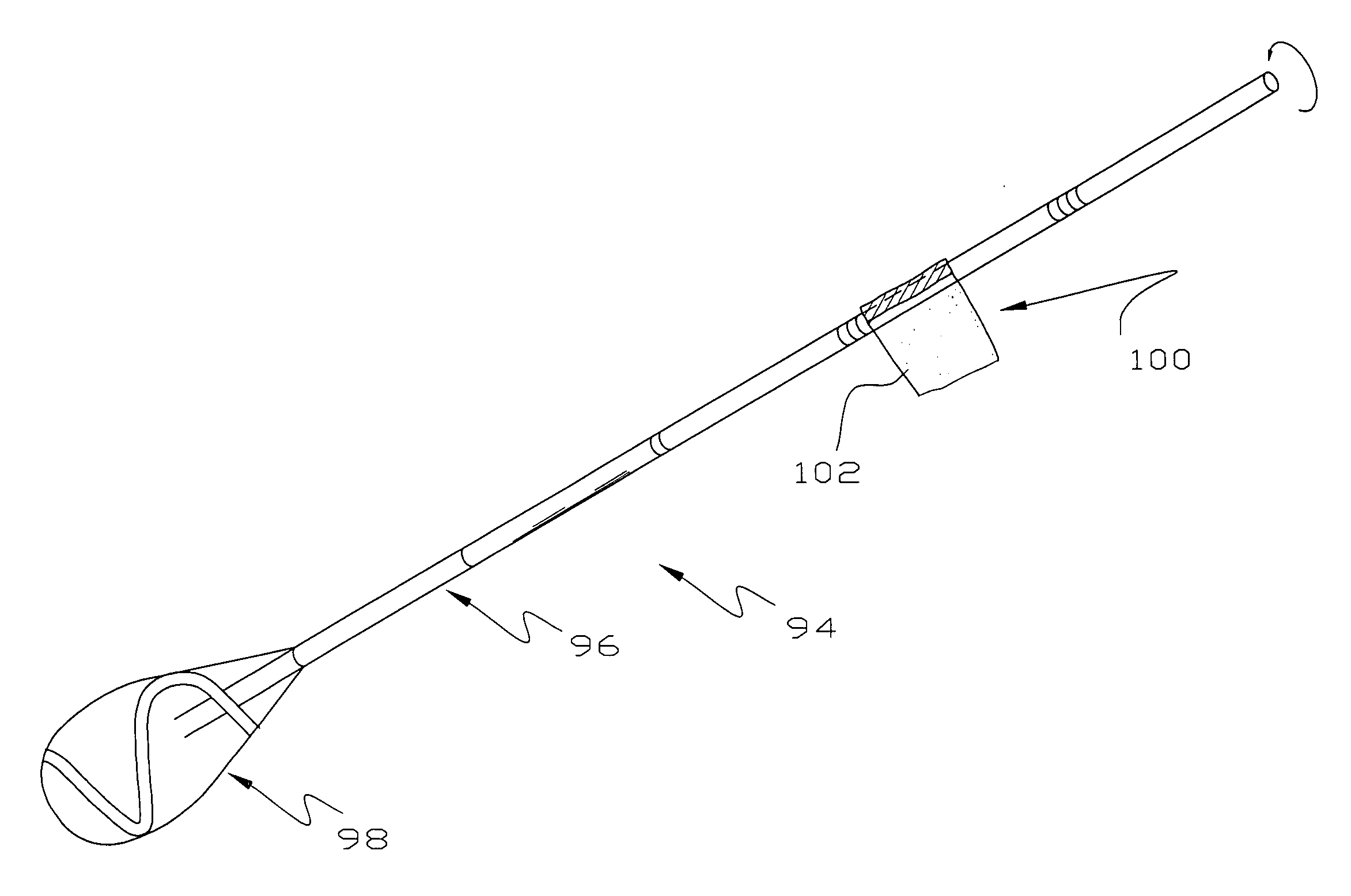
[0010]In this invention, the inside of a cannula of a laparoscopic or thorascopic trocar is cleaned with a swab in the middle of surgery to remove body liquids, tissue or check valve lubricant from the inside of the cannula. Typically, the laparoscope lens becomes clouded and the laparoscope is removed from the cannula and cleaned. Before the laparoscope is reinserted through the cannula, the swab of this invention is run through the cannula to remove any body liquids, tissue or check valve lubricant thereby allowing the lens to generate clear pictures to the monitor being watched by the surgeon.
[0011]The swab is of unusual design and includes a handle of sufficient size to seal against the sealing membrane opening of the cannula to thereby prevent the escape of gas from the patient's abdomen or chest cavity when the swab is being used to clean the inside of the cannula. The swab handle includes depth markings to the end of the sorbent patch on the handle end and / or a shoulder or enlargement at a location where the swab is fully inserted into the cannula. In effect, the enlargement acts as a depth gauge to minimize the potential of dislodging the sorbent patch from the handle and leaving the sorbent patch inside the patient. Radiopaque elements on the sorbent swab end allow the swab end to be located by x-ray or fluoroscope.
[0014]A more specific object of this invention is to provide an improved swab having a handle providing an enlargement to minimize the possibility of the swab being inserted past the end of the cannula.

Problems solved by technology

Occasionally, the interior wall of the cannula collects body liquids, tissue or check valve lubricant, meaning that the lens may become clouded or obscured when reinserted through the cannula.
Although these approaches may theoretically be workable, they have not been accepted, for whatever reasons, by general surgeons in normal every day surgery.

Method used


Image

  • Surgery accessory and method of use
  • Surgery accessory and method of use
  • Surgery accessory and method of use

Examples


Embodiment Construction

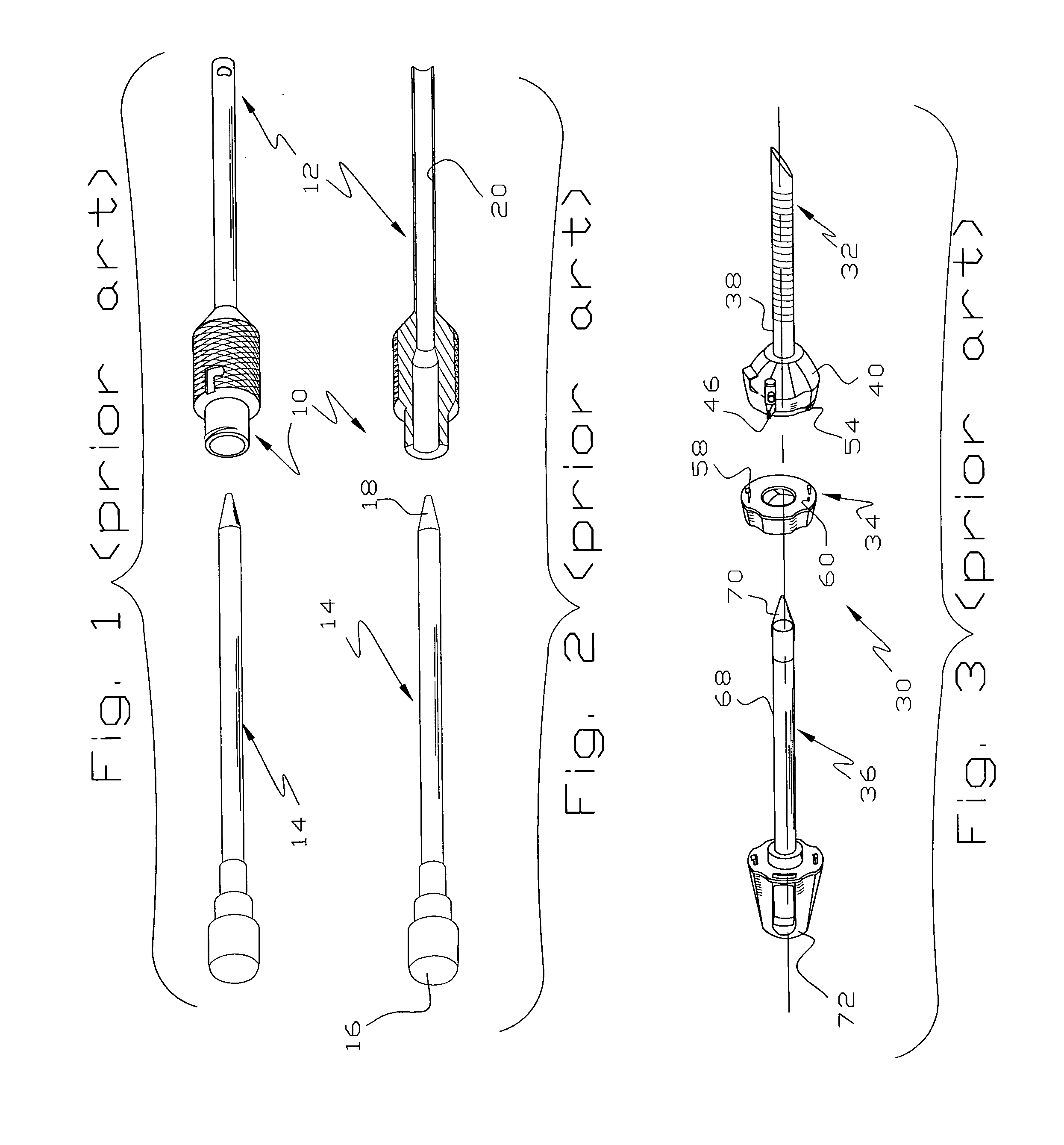
[0023]Referring to FIGS. 1-2, a conventional arthroscopic trocar 10 comprises a cannula 12 into which is inserted a piercing implement or obturator 14. In use, an incision is made in the patient, as on the back of the hand as shown in U.S. Pat. Nos. 5,029,573; 5,318,582 and 5,356,419 and the trocar 10 inserted into the incisions. The trocar 10 is advanced into the patient's body by pushing on an end 16 of the piercing element so the point 18 burrows its way through the patient's flesh to reach the desired location where a surgical procedure is to be conducted. The obturator 14 is then removed, leaving the cannula 12 in place. A series of the trocars 10 may be placed in the patient, depending on the type and extent of surgery to be performed. The arthroscopic trocar 10 will be seen to be quite simple and the cannula 12 is of small internal diameter. Although there may be some variation in the size of the passage 20 through the cannula 12, they are very small and usually are in the 4 ...


PUM


Abstract

In laparoscopic or thorascopic surgery, a trocar is used to provide an passage into the body cavity of a patient for injecting an inert gas to inflate it. A laparoscope or thorascope is passed through a sealing membrane of the trocar so the patient may be observed without releasing a large quantity of the inert gas. If the scope lens is clouded with body liquids or tissue, the scope is removed from the trocar and its lens cleaned. The trocar cannula is cleaned by running a swab through the sealing membrane into the cannula and removing body liquids, tissue and / or check valve lubricant from the inside of the cannula. The swab includes radiopaque markers on the sorbent end, measuring marks on the swab handle and an enlargement on the swab handle. The enlargement is positioned so it may be grasped and the swab pushed into the cannula a distance which is insufficient to push the sorbent end out of the end of the cannula.

Description

[0001]This application is based on Provisional Application Ser. No. 60 / 996,124, filed Nov. 1, 2007 on which priority is claimed.[0002]This invention relates to an accessory for assisting in the performing of laparoscopic or thorascopic surgery and a method of its use and more particularly to a method and apparatus for cleaning an laparoscopic or thorascopic trocar.BACKGROUND OF THE INVENTION[0003]One of the great advances in surgery in the recent past has been the development of remote viewing surgery where the surgeon looks through or uses some type remote viewing instrument to see the site of the procedure. Laparoscopic, thorascopic, arthroscopic or endoscopic surgery are species of remote viewing surgery and involve the introduction of a viewing scope into the interior of a patient where a surgical procedure is to be performed. In the case of endoscopic surgery, an endoscope is inserted through a natural body opening. In the case of arthroscopic surgery, an incision is made near ...

Claims


Application Information

Patent Timeline
Patent Type & Authority Applications(United States)
IPC IPC(8): A61B17/34B08B9/04A61M31/00
CPCA61B17/3421A61B19/34A61B19/54B08B9/00A61M2025/0019B08B1/00A61B2019/4857A61B90/70A61B2090/0811A61B90/39A61B17/3462B08B1/145
Inventor GRIFFIN, GLENN A.
Owner GRIFFIN GLENN A
Why Eureka
Eureka Blog
Learn More