Non-Invasive In Vivo Imaging and Methods for Treating Type I Diabetes
- Summary
- Abstract
- Description
- Claims
- Application Information
AI Technical Summary
Benefits of technology
Problems solved by technology
Method used
Image
Examples
example 1
REFERENCES FOR EXAMPLE 1
[0052]1. Wajchenberg, B. L. Beta-Cell Failure in Diabetes and Preservation by Clinical Treatment. Endocr Rev (2007).[0053]2. Berggren, P. O. & Leibiger, I. B. Novel aspects on signal-transduction in the pancreatic beta-cell. Nutr Metab Cardiovasc Dis 16 Suppl 1, S7-10 (2006).[0054]3. Vetterlein, F., Petho, A. & Schmidt, G. Morphometric investigation of the microvascular system of pancreatic exocrine and endocrine tissue in the rat. Microvasc Res 34, 231-8 (1987).[0055]4. Woods, S. C. & Porte, D., Jr. Neural control of the endocrine pancreas. Physiol Rev 54, 596-619 (1974).[0056]5. Rahier, J., Goebbels, R. M. & Henquin, J. C. Cellular composition of the human diabetic pancreas. Diabetologia 24, 366-71 (1983).[0057]6. Köhler, M. et al. Imaging of Pancreatic Beta-Cell Signal-Transduction. Curr. Med. Chem.-Immun., Endoc. &Metab. Agents 4, 281-299 (2004).[0058]7. Speier, S. & Rupnik, M. A novel approach to in situ characterization of pancreatic beta-cells. Pfluger...
example 2
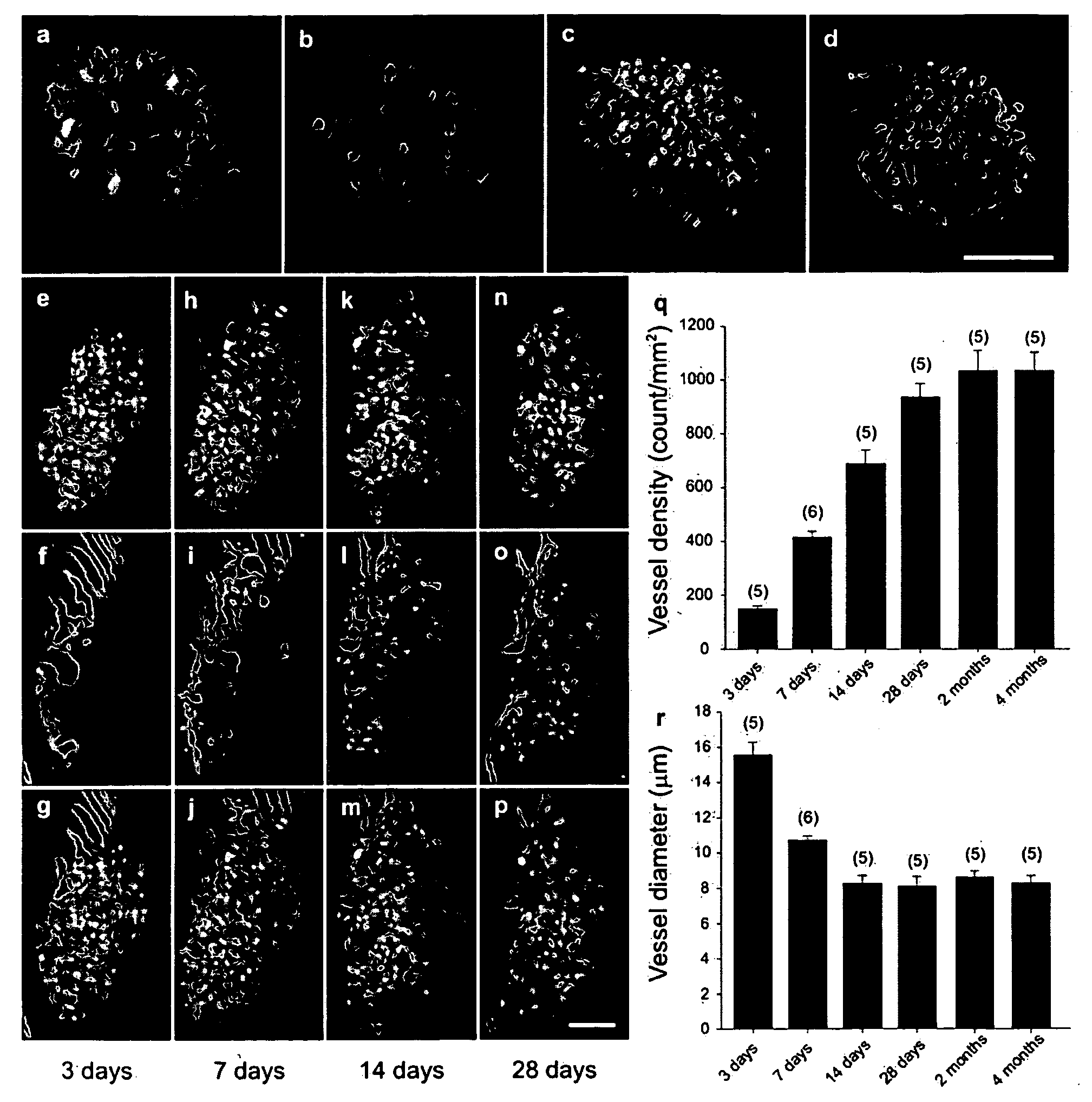
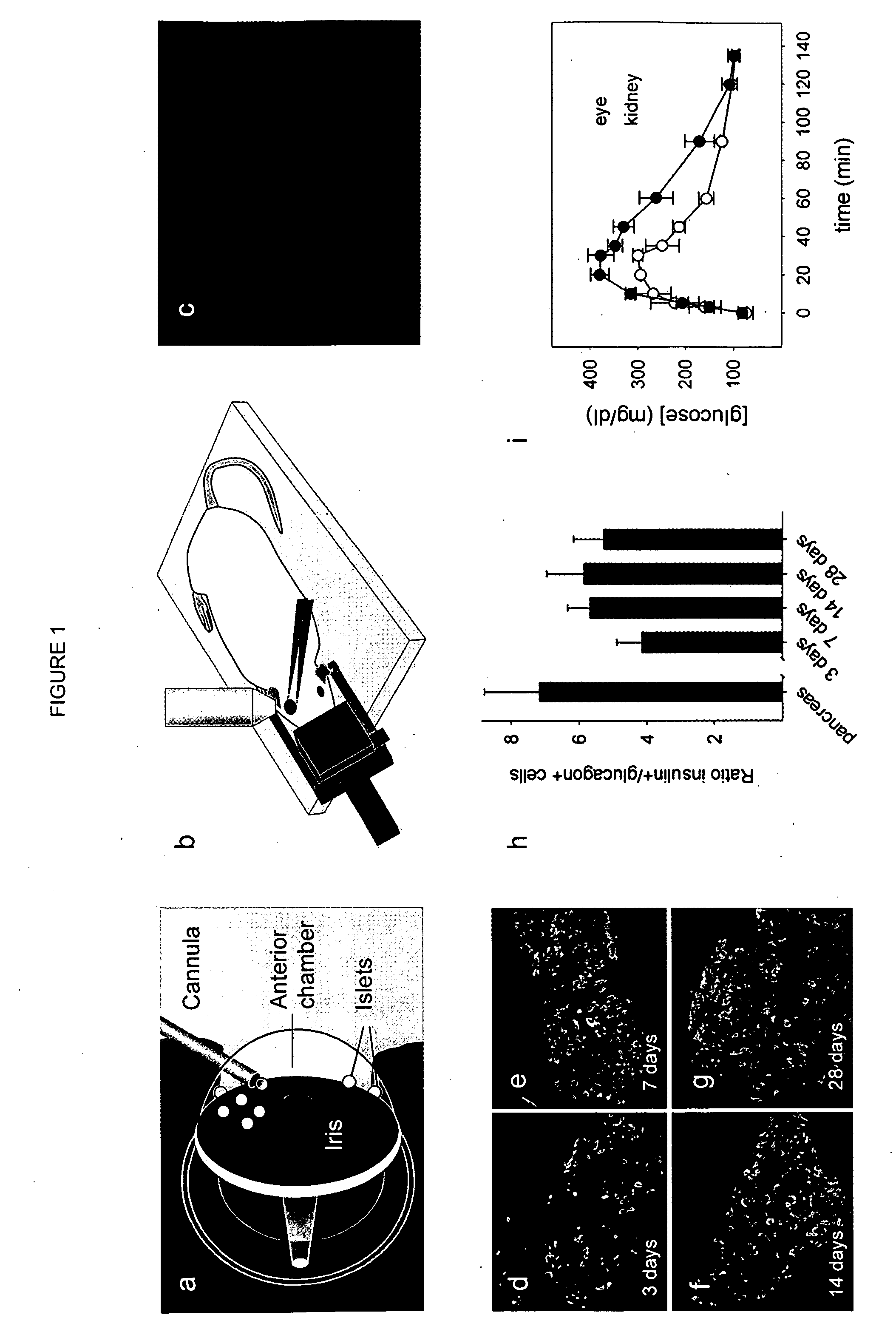
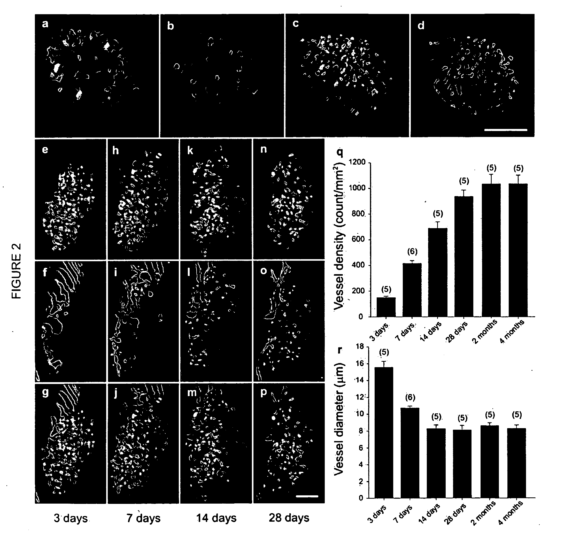
[0080]In this example, we provide a step-by-step protocol for non-invasive longitudinal in vivo studies of cell biology at single-cell resolution, taking advantage of the cornea as a natural body window. For this purpose, the tissue of interest is transplanted into the anterior chamber of the eye and cell biological parameters are assessed by LSM through the cornea. The anterior chamber of the eye has been frequently used as a transplantation site to study a variety of tissues3-7. While originally the anterior chamber of the eye was selected as a transplantation site because of its properties as an immune privileged site8, most studies utilized the anterior chamber in a syngeneic transplantation setting because it is easily accessible and the cornea allows macroscopic observation of the engrafted tissue. Additionally, the iris, which forms the base of the anterior chamber, has one of the highest concentrations of blood vessels and autonomic nerves in the body, and thereby enables fa...
example 2 references
[0230]1. Koo, V., Hamilton, P. W. & Williamson, K. Non-invasive in vivo imaging in small animal research. Cell Oncol 28, 127-139 (2006).[0231]2. Handbook of biological confocal microscopy, Edn. 3 (Pawley, J. B.) (Springer, New York, N.Y., 2005).[0232]3. Adeghate, E., Ponery, A. S., Ahmed, I. & Donath, T. Comparative morphology and biochemistry of pancreatic tissue fragments transplanted into the anterior eye chamber and subcutaneous regions of the rat. European journal of morphology 39, 257-268 (2001).[0233]4. Katoh, N., et al. Target-specific innervation by autonomic and sensory nerve fibers in hairy fetal skin transplanted into the anterior eye chamber of adult rat. Cell and tissue research 266, 259-263 (1991).[0234]5. Olson, L. & Seiger, A. Beating intraocular hearts: light-controlled rate by autonomic innervation from host iris. Journal of neurobiology 7, 193-203 (1976).[0235]6. Wu, W., Scott, D. E. & Reiter, R. J. Transplantation of the mammalian pineal gland: studies of surviv...
PUM
 Abstract
Description
Claims
Application Information
 - R&D
- Intellectual Property
- Life Sciences
- Materials
- Tech Scout
- Unparalleled Data Quality
- Higher Quality Content
- 60% Fewer Hallucinations
Browse by: Latest US Patents, China's latest patents, Technical Efficacy Thesaurus, Application Domain, Technology Topic, Popular Technical Reports.
© 2025 PatSnap. All rights reserved.Legal|Privacy policy|Modern Slavery Act Transparency Statement|Sitemap|About US| Contact US: help@patsnap.com


