Methods for Intraoperative Organotypic Nerve Mapping

a technology of organotypic nerve and intraoperative operation, which is applied in the field of organotypic nerve mapping methods, can solve the problems of inability to distinguish penile, urethral, or prostatic nerves, and the current use of imaging technology during surgery is generally limited to ultrasound, pet, open mri or other expensive, cumbersome devices,

Inactive Publication Date: 2008-08-14
UNIV OF VIRGINIA ALUMNI PATENTS FOUND
View PDF1 Cites 23 Cited by
  • Summary
  • Abstract
  • Description
  • Claims
  • Application Information

AI Technical Summary

Benefits of technology

[0006]The present invention provides a real time method for visualizing nerves in vivo. In one aspect, the invention provides for visualizing cavernous nerves in vivo. The present invention further provides a method to aid surgeons in nerve preservation and tumor border identification during robotic radical prostatectomy and during other surgical procedures where it is important to identify and preserve nerves.
[0007]Because of the recent commercial availability of portable, fluorescent, fiber-optic laser confocal microscopes, it is now practical to employ the methods of the present invention for acquiring repeated microscopic images of living tissues of interest with minimal damage to these tissues. The unique high-sensitivity design of the present invention allows imaging of structures as small and as difficult to locate as the fine autonomic nerves fibers to internal organs. In one aspect, when used intra-operatively with the da Vinci robotic platform to simultaneously identify cavernous nerves (to avoid injury resulting in erectile dysfunction (ED)) and prostate cancer (to prevent the occurrence of positive tumor margins); it is envisioned that this technique will revolutionize surgery, allowing the possibility of molecular and cellular surgery rather than gross anatomical surgery, thereby reducing cancer recurrences and reducing morbidity associated with surgery.
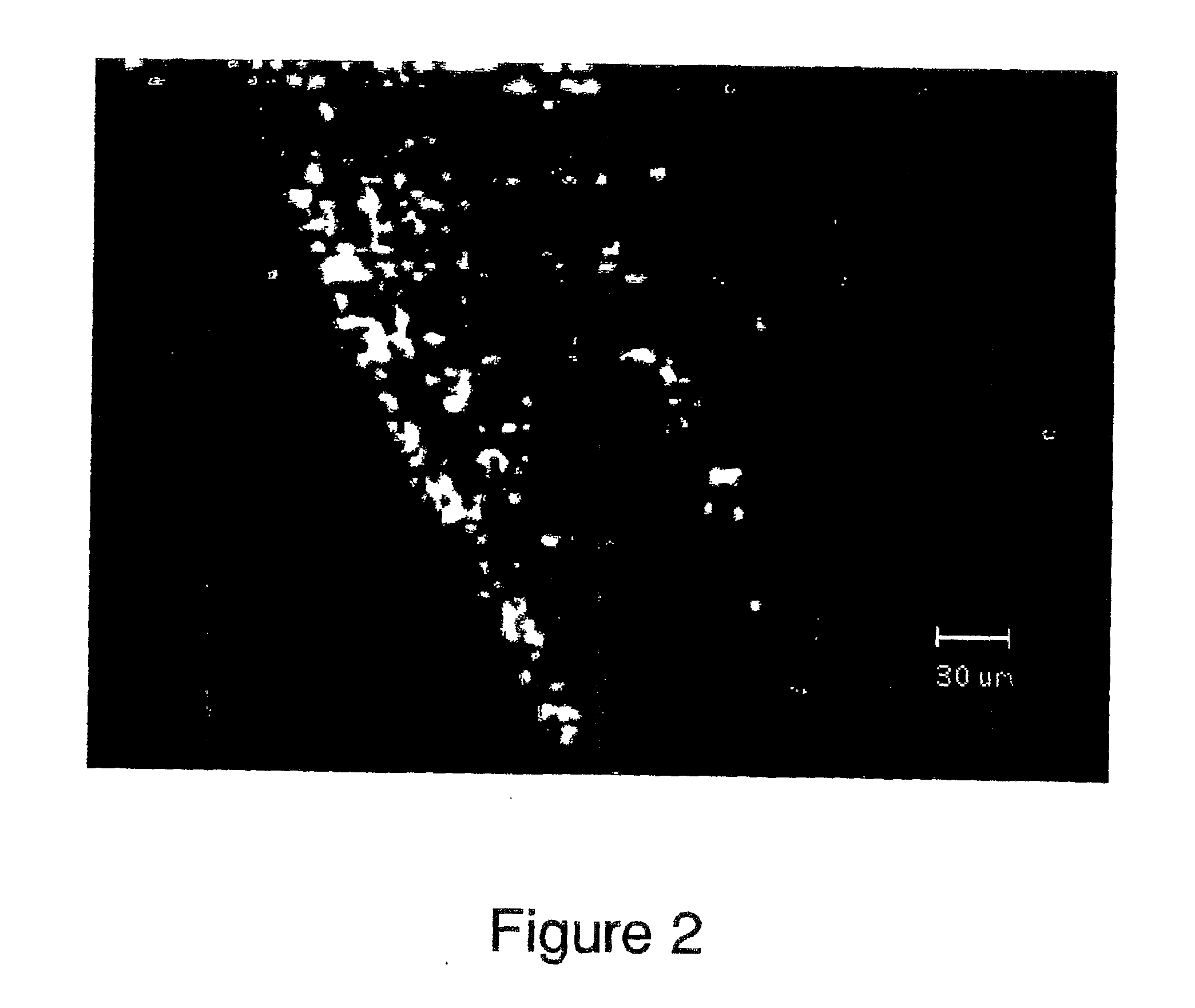
[0012]Clinically, in one aspect, the present invention encompasses injection of tracer into the penis at about 24 hours prior to surgery. In one aspect, one or both corpora cavernosa are injected. In another aspect, one or both crura of the penis are injected. In yet another aspect, the corpus spongiosum is injected. In a further aspect, other tissues in the area are injected. It will be appreciated by one of ordinary skill in the art that combinations of these sites could also be injected. The invention further encompasses injecting various amounts of tracer, as well as injecting at different amounts of time prior to surgery. Then, during robotic-assisted laparoscopic prostatectomy or cystectomy the Cell Vizio device would be mounted on a robotic stabilizer (arm) and brushed along side the neurovascular bundle during dissection to provide a road map to safely avoid cauterizing or applying traction on these fine nerve fibers. The novelty of the technique is derived from: 1) the use of retrograde tracers to positively identify exactly those nerves (axons as opposed to cell bodies) supplying the penis that control penile erection that course beside the prostate; and 2) employing fiber optic laser confocal fluorescence microscopy to non-invasively, reversibly image these nerves intra-operatively. This process allows viewing labeled axons and not connective or vascular tissues that obscure the nerves without altering the function of the cavernous nerves. This unique method of labeling and imaging during surgery makes it possible for us to detect the nerves at various levels during the surgery, thus avoid damaging them.

Problems solved by technology

Current use of imaging technology during surgery is generally limited to ultrasound, PET, open MRI or other expensive, cumbersome devices that lack adequate cellular resolution for fine tumor border localization and organ specific nerve identification.
Optical imaging on the cellular level has been restricted to in vitro applications due to problems with optical access or labeling.
Some investigators have tried staining for NADPH found in nerves to the penis although this methodology fails to distinguish penile, urethral, or prostatic nerves.

Method used


Image

  • Methods for Intraoperative Organotypic Nerve Mapping
  • Methods for Intraoperative Organotypic Nerve Mapping
  • Methods for Intraoperative Organotypic Nerve Mapping

Examples


examples

[0131]The invention is now described with reference to the following examples. These examples are provided for the purpose of illustration only and the invention should in no way be construed as being limited to these examples but rather should be construed to encompass any and all variations which become evident as a result of the teaching provided herein.

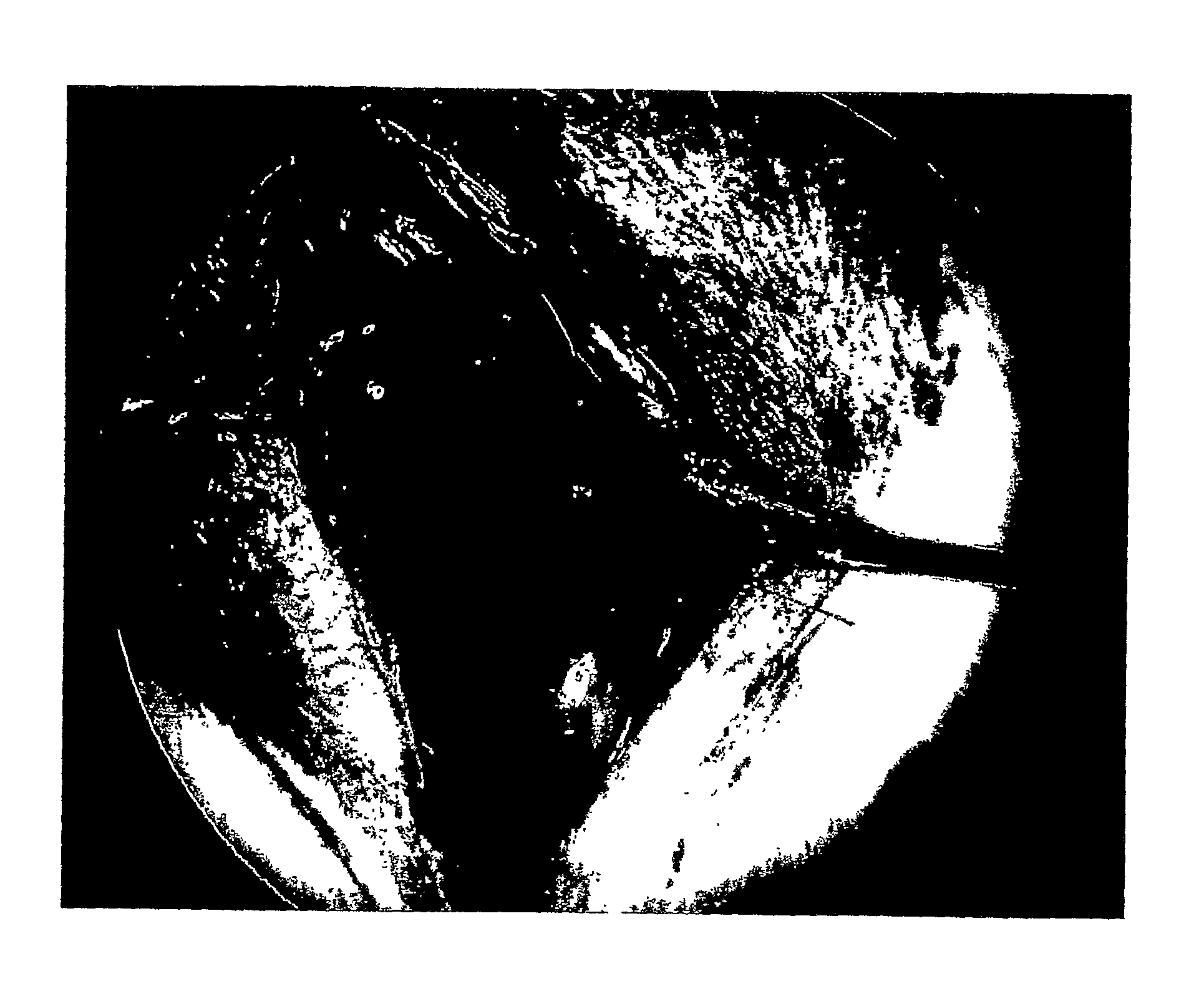
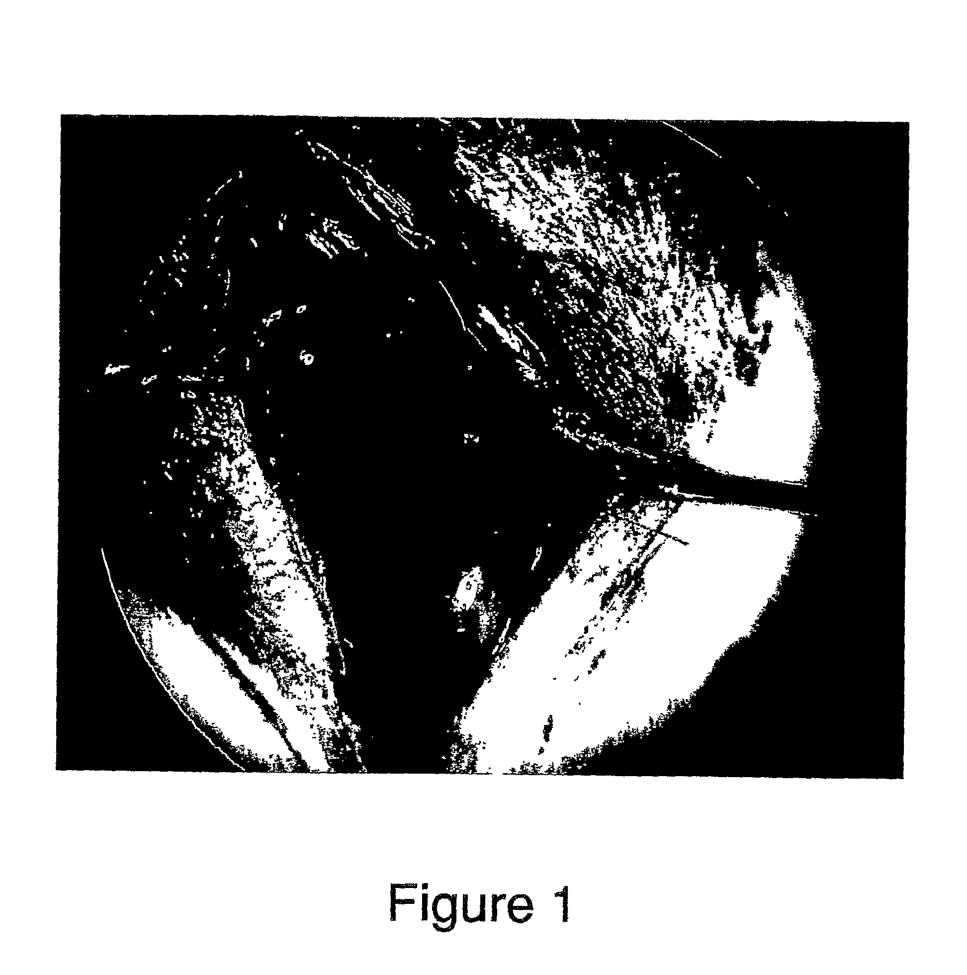
[0132]The present invention provides composition, methods, and apparatuses for visualizing the cavernous nerves that directly control erectile function in real time in a living subject. The technique was developed to aid surgeons in nerve preservation during procedures, including, but not limited to, robotic radical prostatectomy, by allowing them to positively identify the anatomical position of the nerves in real time.

[0133]A feasibility study of intraoperative cavernous nerve imaging was performed. The aim of the study was to evaluate the novel nerve imaging protocol of the invention for its utility as a real-time surgical aid....


PUM

PropertyMeasurementUnit
fluorescence lifetimeaaaaaaaaaa
w/waaaaaaaaaa
diametersaaaaaaaaaa

Abstract

The present invention relates to compositions, methods and apparatuses for locating a nerve during a surgical procedure.

Description

CROSS REFERENCE TO RELATED APPLICATIONS[0001]This application is entitled to priority pursuant to 35 U.S.C. §119(e) to U.S. provisional patent application No. 60 / 709,872, filed on Aug. 19, 2005, the entirety of which is incorporated by reference herein.FIELD OF THE INVENTION[0002]The present invention relates to compositions, methods, and apparatuses for locating a nerve during a surgical procedure. The invention also relates to preserving a nerve from injury during a surgical procedure.BACKGROUND[0003]Studies have shown that as many as two thirds of men develop erectile dysfunction (ED) following radical prostatectomy for prostate cancer or radical cystectomy for bladder cancer as a result cavernous nerve injury during surgery. The cavernous nerves are small autonomic nerve fibers that course along the capsule of the prostate to the corpus cavernosum of the penis. These nerves are responsible for penile erection. Potency rates (i.e., cavernous nerve preservation) vary dramatically ...

Claims


Application Information

Patent Timeline
Patent Type & Authority Applications(United States)
IPC IPC(8): A61B5/04
CPCA61B5/0059A61B5/4393A61K49/0056A61K47/48261A61K49/0041A61B5/4893A61K47/6415
Inventor STEERS, WILLIAM D.BOYETTE, LISA BLACKBURN
Owner UNIV OF VIRGINIA ALUMNI PATENTS FOUND
Why Eureka
Try Eureka