
System and Method for Automatic Detection of Rib Metastasis in Computed Tomography Volume

a computed tomography and volume technology, applied in image analysis, instruments, computing, etc., can solve the problems of easy error and tedious diagnostic process

Active Publication Date: 2008-06-12
SIEMENS MEDICAL SOLUTIONS USA INC
View PDF14 Cites 20 Cited by
  • Summary
  • Abstract
  • Description
  • Claims
  • Application Information

AI Technical Summary

Problems solved by technology

This diagnosis process is tedious and prone to error since the ribs typically cross through the axial slices at oblique angles.

Method used


Image

  • System and Method for Automatic Detection of Rib Metastasis in Computed Tomography Volume
  • System and Method for Automatic Detection of Rib Metastasis in Computed Tomography Volume
  • System and Method for Automatic Detection of Rib Metastasis in Computed Tomography Volume

Examples


Embodiment Construction

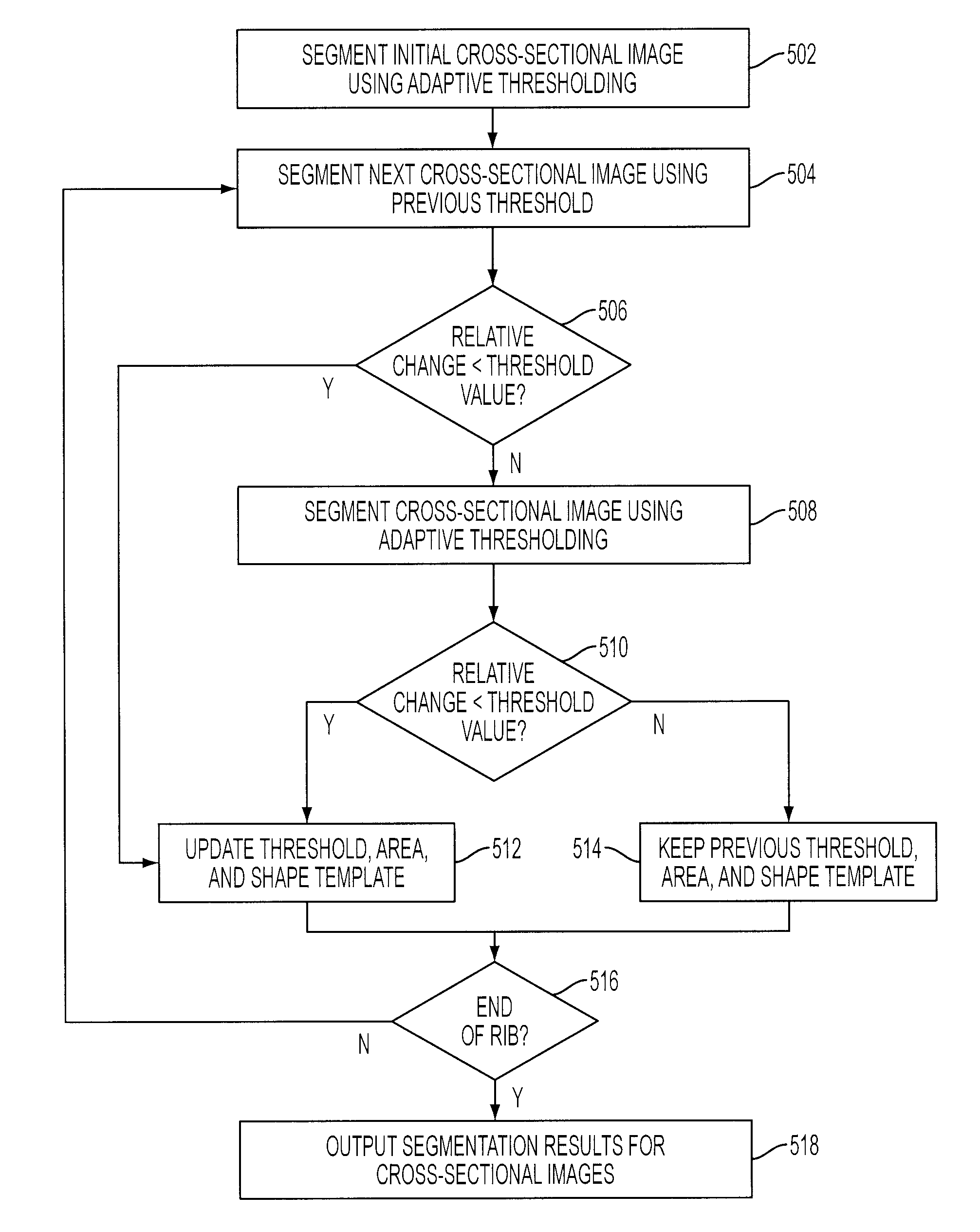
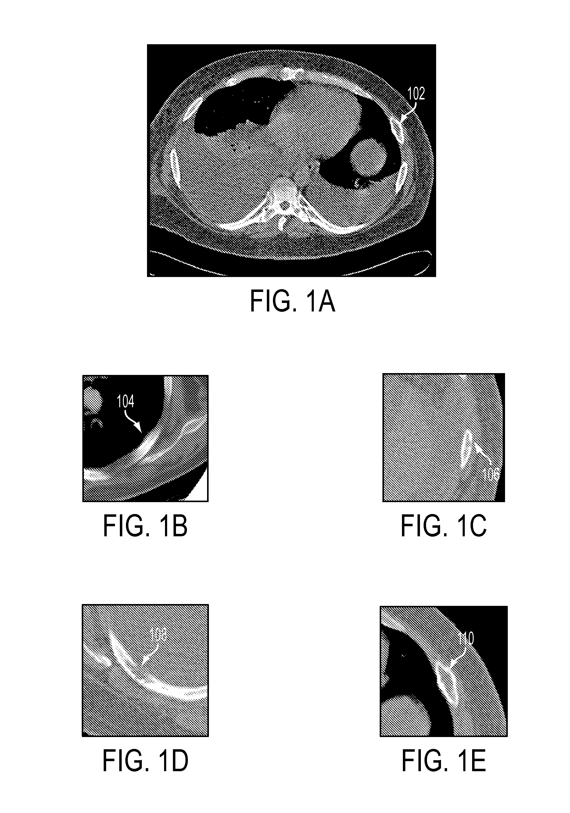
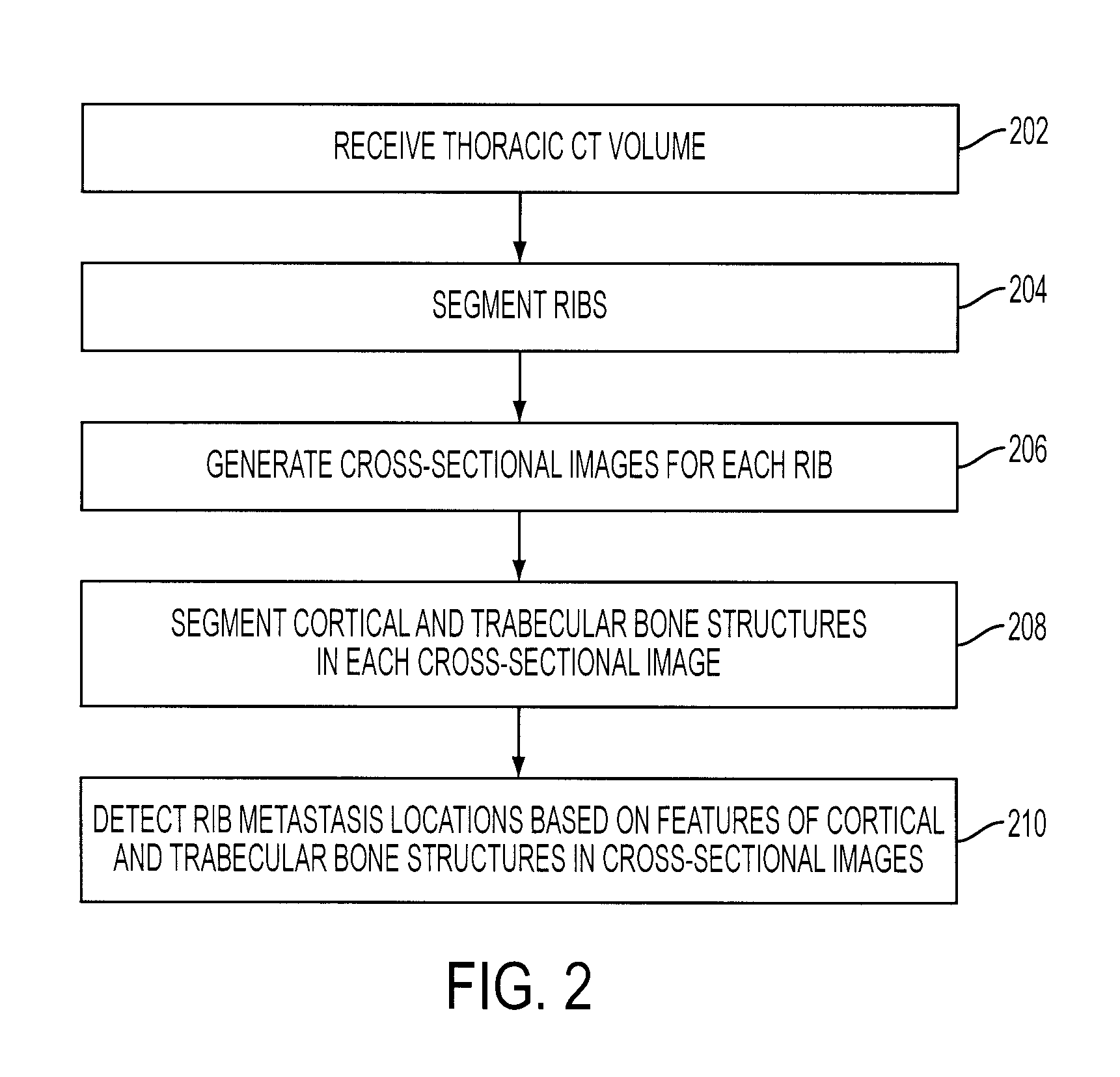
[0017]The present invention is directed to a method for automatically detecting rib metastasis in computed tomography (CT) volumes. Embodiments of the present invention are described herein to give a visual understanding of the rib metastasis detection method. A digital image is often composed of digital representations of one or more objects (or shapes). The digital representation of an object is often described herein in terms of identifying and manipulating the objects. Such manipulations are virtual manipulations accomplished in the memory or other circuitry / hardware of a computer system. Accordingly, is to be understood that embodiments of the present invention may be performed within a computer system using data stored within the computer system. For example, according to various embodiments of the present invention, electronic data representing a thoracic CT volume is manipulated within a computer system in order to detect rib metastasis in the thoracic CT volume.

[0018]Accord...


PUM


Abstract

A method and system for automatically detecting rib metastasis in a thoracic CT volume is disclosed. The ribs are segmented in said CT volume by recursive tracing. A series of cross-sectional images are then generated along a centerline of each rib. Cortical and trabecular bone structures are segmented in each of the cross-sectional images for each rib. Features are calculated for each cross-sectional image based on characteristics of the cortical and trabecular bone structures, and alterations are detected in the cross-sectional images based on the features. Rib metastasis is detected in a rib when an alteration is detected in a number of consecutive cross-sectional images along the centerline of the rib.

Description

[0001]This application claims the benefit of U.S. Provisional Application No. 60 / 869,354, filed Dec. 11, 2006, the disclosure of which is herein incorporated by reference.BACKGROUND OF THE INVENTION[0002]The present invention relates to detecting rib metastasis in computed tomography (CT) volumes, and more particularly to a system and method for automatic detection of rib metastasis in CT volumes.[0003]Computed tomography (CT) is a medical imaging method whereby digital geometry processing is used to generate a three-dimensional image of the internal features of a patient from a large series of two-dimensional X-ray images taken around a single axis of rotation. Such CT imaging results in CT volume data which is a virtual representation of internal anatomical features of a patient. The CT volume data consists of multiple slices, or two-dimensional images, that can be combined to generate a three dimensional image. CT imaging is particularly useful because it can show several types o...

Claims


Application Information

Patent Timeline
Patent Type & Authority Applications(United States)
IPC IPC(8): G06K9/00
CPCG06T7/0012
Inventor SHEN, HONGMA, LIMIN
Owner SIEMENS MEDICAL SOLUTIONS USA INC
Learn More