Eureka AIR delivers breakthrough ideas for toughest innovation challenges, trusted by R&D personnel around the world.

Therapeutic Administration Of The Scrambled Anti-Angiogenic Peptide C16Y

a technology of anti-angiogenic peptides and scrambled proteins, which is applied in the field of ocular disease, can solve problems such as vision loss

Inactive Publication Date: 2008-06-05
DEPT OF HEALTH & HUMAN SERVICES SEC OF GOVERNMENT OF THE UNITED STATES OF AMERICA AS REPRESENTED BY THE
View PDF2 Cites 5 Cited by
  • Summary
  • Abstract
  • Description
  • Claims
  • Application Information

AI Technical Summary

Benefits of technology

[0012]Angiogenesis plays a key role in several pathological conditions, including cancer and ocular neovascularization. Tumors are unable to grow and metastasize without the formation of new blood vessels. Angiogenesis in the eye (ocular neovascularization) is associated with a variety of ocular diseases, including age-related macular degeneration (AMD) and diabe

Problems solved by technology

Corneal neovascularization, often associated with the use of contact lenses, can lead to vision loss.

Method used


Image

  • Therapeutic Administration Of The Scrambled Anti-Angiogenic Peptide C16Y
  • Therapeutic Administration Of The Scrambled Anti-Angiogenic Peptide C16Y
  • Therapeutic Administration Of The Scrambled Anti-Angiogenic Peptide C16Y

Examples


example 1

Endothelial Cell Adhesion by C16 and Scrambled C16 Peptides

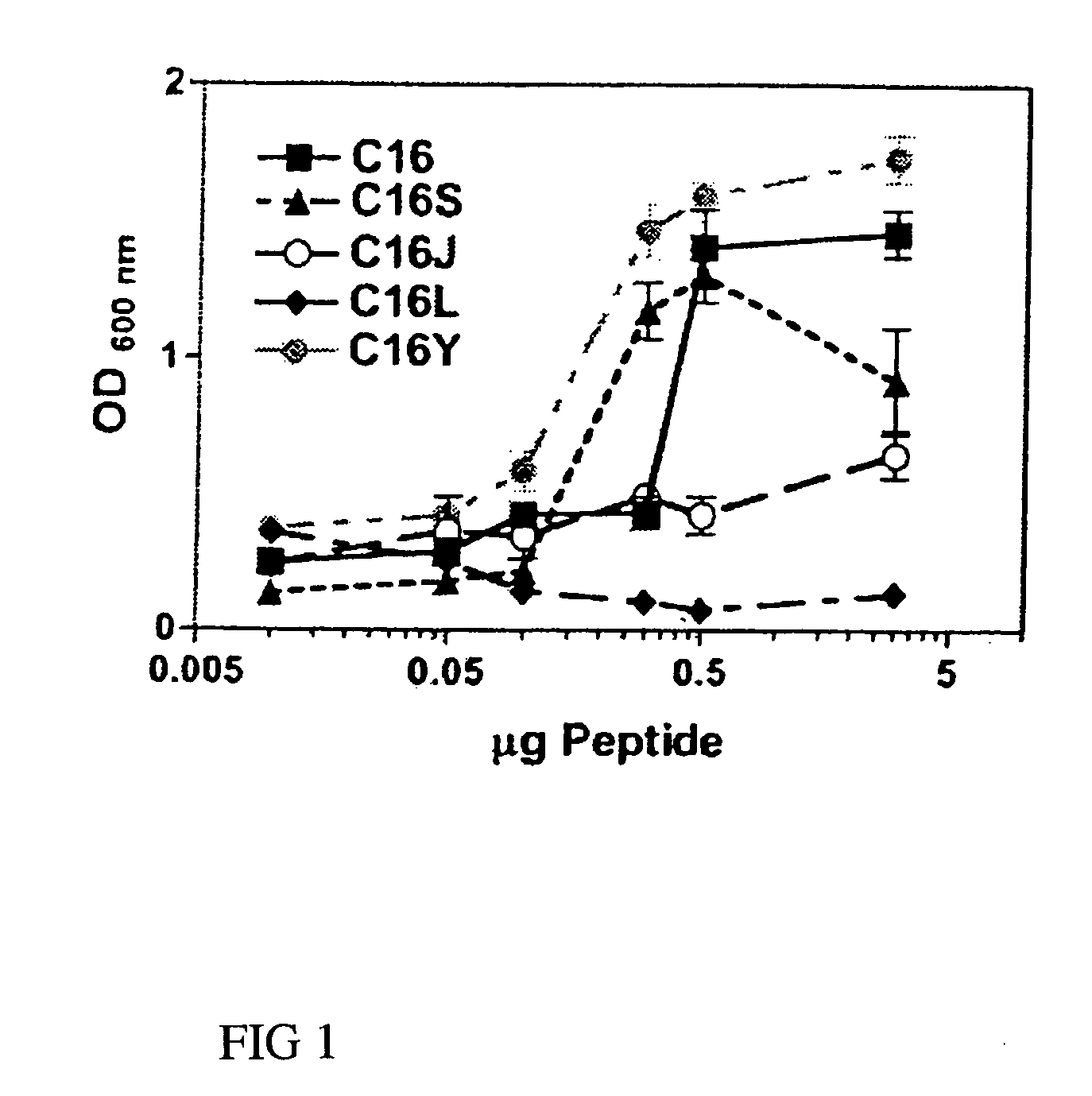
[0055]Endothelial cell adhesion assays were performed on 96-well plates coated overnight with either laminin-1 (0.5 μg) or synthetic laminin peptides C16, C16S, C16J, C16L, or C16Y (0-5 μg) as described previously (Malinda 1999). Wells were rinsed with PBS, blocked with 2 mg / ml of BSA, and rinsed again with PBS. Cell adhesion was performed using 0.1 ml of RPMI 1640 containing 35,000 HUVECs. After a 1.5-hour incubation period at 37° C., unbound cells were decanted and attached cells were fixed and stained with 20% methanol / 0.2% crystal violet. Dishes were extensively rinsed, and bound dye was solubilized in 2% SDS and quantitated at 600 nm. Assays were done in triplicate at least three times.

[0056]Cells attached to C16, the parent peptide, and to C16S at amounts >0.1 μg (FIG. 1). Although C16Y showed a similar attachment pattern to that of C16 and C16S, more cells adhered to it than to any other peptide at all of the amounts ...

example 2

Ability of C16 and Scrambled C16 Peptides to Block Laminin-1 Endothelial Cell Adhesion

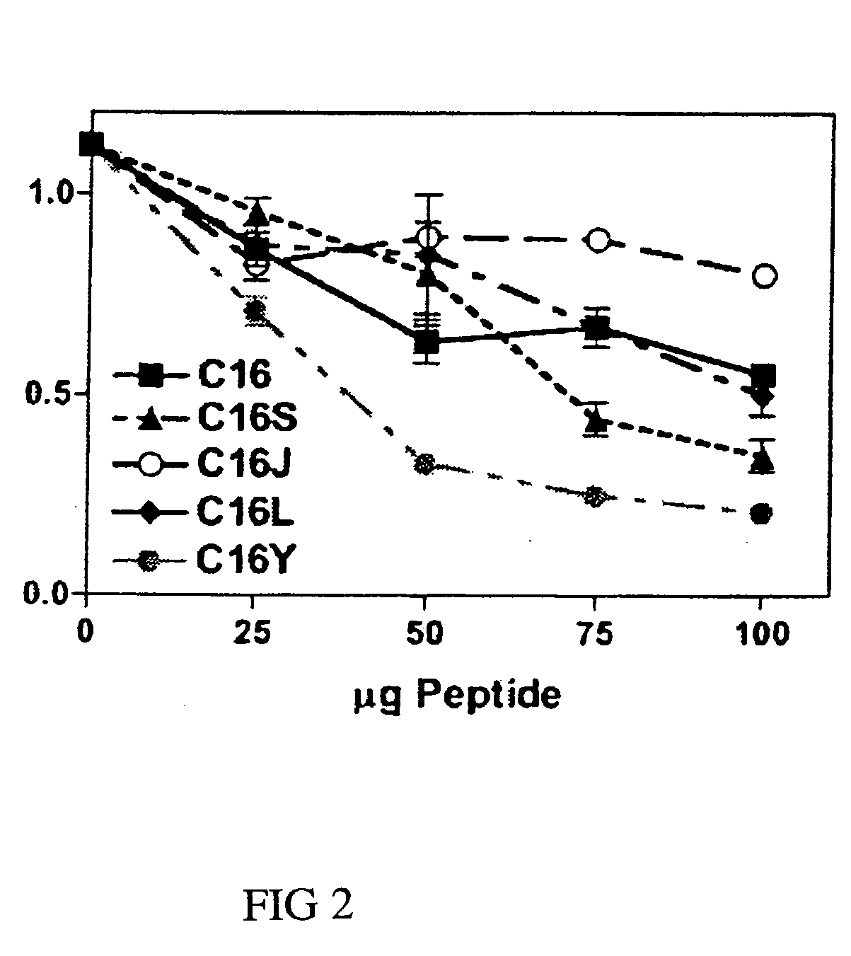
[0057]Competition experiments were done using 96-well plates coated with laminin-1 (0.5 μg) in the presence of 0-100 μg / ml of competing C16, C16S, C16J, C16L, or C16Y synthetic laminin peptide as specified. Control wells were coated with BSA only. Wells were contacted with 0.1 ml of RPMI 1640 containing 35,000 HUVECs. After a 1.5-hour incubation at 37° C., unbound cells were decanted and attached cells were fixed and stained with 20% methanol / 0.2% crystal violet. Dishes were extensively rinsed, and bound dye was solubilized in 2% SDS and quantitated at 600 nm. Assays were done in triplicate at least three times.

[0058]C16Y had the strongest inhibitory activity at all concentrations tested (FIG. 2). The most significant differences were observed at 50 μg / ml, where C16Y inhibited endothelial cell attachment to laminin-1 by >70%. C16 and C16S, on the other hand, inhibited by 75 μg / ml.

example 3

Disruption of Endothelial Cell Tube Formation by C16Y

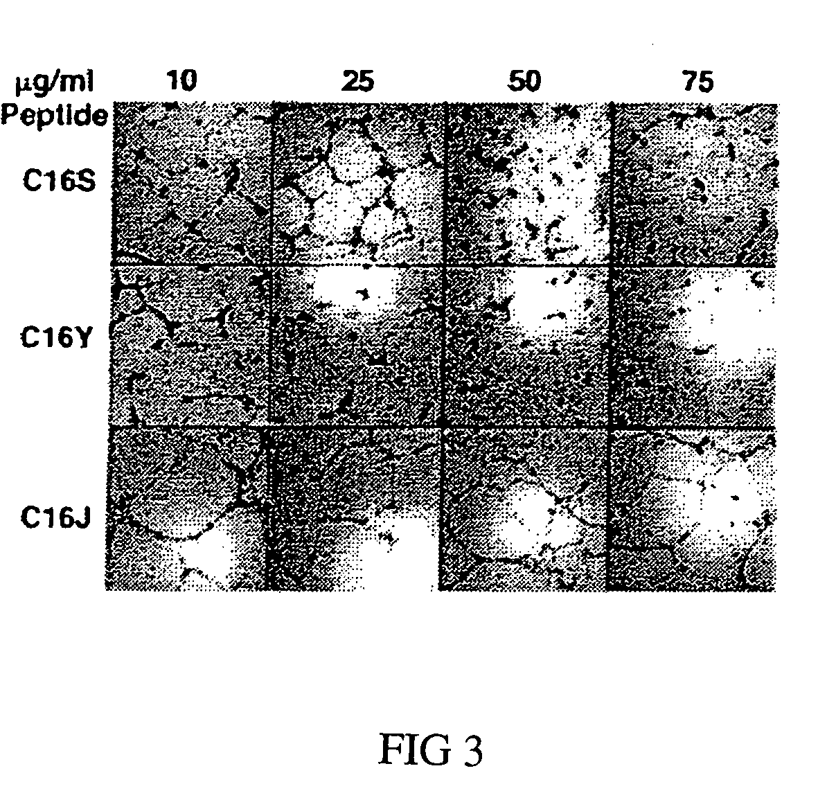
[0059]Tube-forming assays were performed as described previously (Ponce 2001). 48-well plates were coated with 200 μl / well of Matrigel (Gho 2001; Kubota 1988), a basement membrane matrix. HUVECs (24,000 cells / well) were plated in RPMI 1640 containing 10% bovine calf serum, defined and supplemented (HyClone Laboratories, Inc., Logan, Utah), along with 100 mg / liter of endothelial cell growth factor (Collaborative Biomedical). C16 or scrambled C16 peptides were added at concentrations ranging from 10-75 μg / ml. Control wells contained medium only. After 16 hours, cells were fixed and stained with Diff-Quick fixative (methanol) and solution II (6.25% (w / v) each of azure A and methylene blue) (Dade A G, Dudingen, Switzerland), and tube formation was scored by a blinded observer. Each peptide was tested at least in triplicate, and the assays were repeated a minimum of three times.

[0060]As expected, C16 and C16S disrupted tube formation a...


PUM

PropertyMeasurementUnit
Compositionaaaaaaaaaa

Abstract

Unregulated angiogenesis is associated with a variety of pathological conditions. Tumor growth and metastasis is dependent on the development of new blood vessels. The development of new blood vessels in the eye, or ocular neovascularization, has been implicated in a variety of serious ocular diseases. For instance, choroidal neovascularization is linked to age-related macular degeneration, while retinal neovascularization is linked to diabetic retinopathy. The present invention is based on the discovery of a peptide sequence, C16Y, which inhibits ocular neovascularization and tumor growth in vivo. C16Y is a scrambled version of the C16 peptide sequence from the y1 chain of laminin-1. Unlike C16, which is an angiogenic stimulator, C16Y has been shown to inhibit angiogenesis. The present invention discloses methods of treating ocular neovascularization and cancer using both full-length and truncated versions of the C16Y.

Description

FIELD OF THE INVENTION[0001]The present invention relates to the field of ocular disease, in particular diseases associated with ocular neovascularization, and to the field of cancer therapy, specifically breast cancer therapy. In addition, the present invention relates to angiogenesis, angiogenesis inhibitors, integrins, and integrin pathway inhibition.BACKGROUND[0002]The extracellular matrix (ECM), often referred to as connective tissue, is the complex structure that surrounds and supports cells in mammalian tissue. It is composed primarily of three classes of biomolecules: structural proteins (collagen and elastin), specialized proteins (e.g., laminin, fibronectin, fibrillin), and proteoglycans (core proteins linked to repeating disaccharides known as glycosaminoglycans or GAGs) (Pepper 2001). The ECM is vital for the maintenance and differentiation of many cell types, including the endothelium. In addition, it plays a role in the formation of new blood vessels from pre-existing ...

Claims


Application Information

Patent Timeline
IPC IPC(8): A61K39/44A61K38/10A61P35/00A61P27/02A61K38/08A61K38/00C07K5/103C07K5/107C07K14/78
CPCA61K38/08A61K38/10C07K14/78C07K5/1016C07K5/101A61P27/02A61P27/06A61P35/00A61P9/00
Inventor CSAKY, KARL G.KLEINMAN, HYNDAPONCE, LOURDES
Owner DEPT OF HEALTH & HUMAN SERVICES SEC OF GOVERNMENT OF THE UNITED STATES OF AMERICA AS REPRESENTED BY THE
Why Eureka
Eureka Blog
Learn More