Composition and Methods for Promoting Wound Healing and tissue Regeneration
a technology of tissue regeneration and composition, applied in the direction of peptide/protein ingredients, dna/rna fragmentation, depsipeptides, etc., can solve the problems of complex tissue structure and function, rarely, if ever wholly restored, scarring, etc., and achieve the effect of promoting wound healing
- Summary
- Abstract
- Description
- Claims
- Application Information
AI Technical Summary
Benefits of technology
Problems solved by technology
Method used
Image
Examples
example 1
In Vitro Scratch Injury
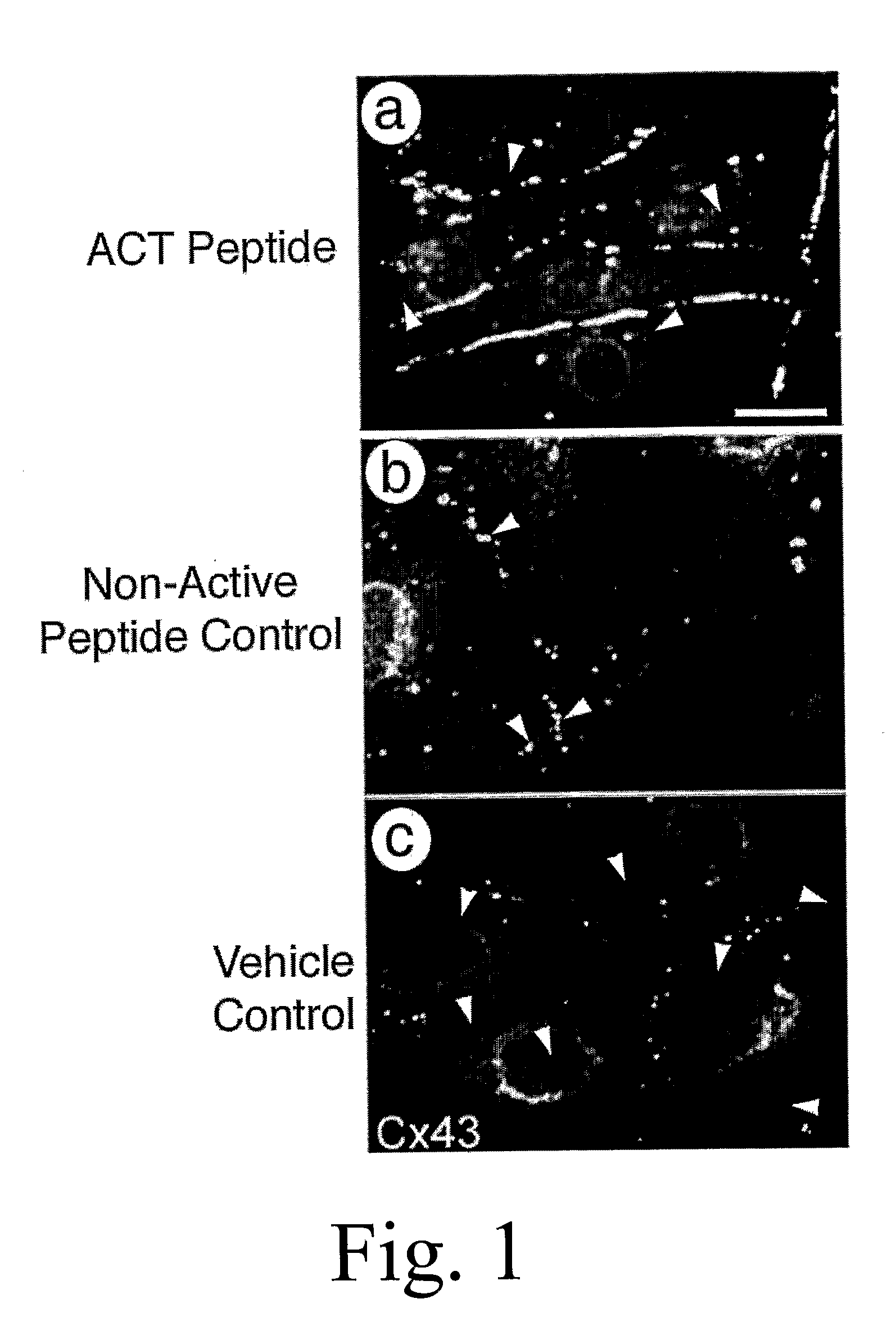
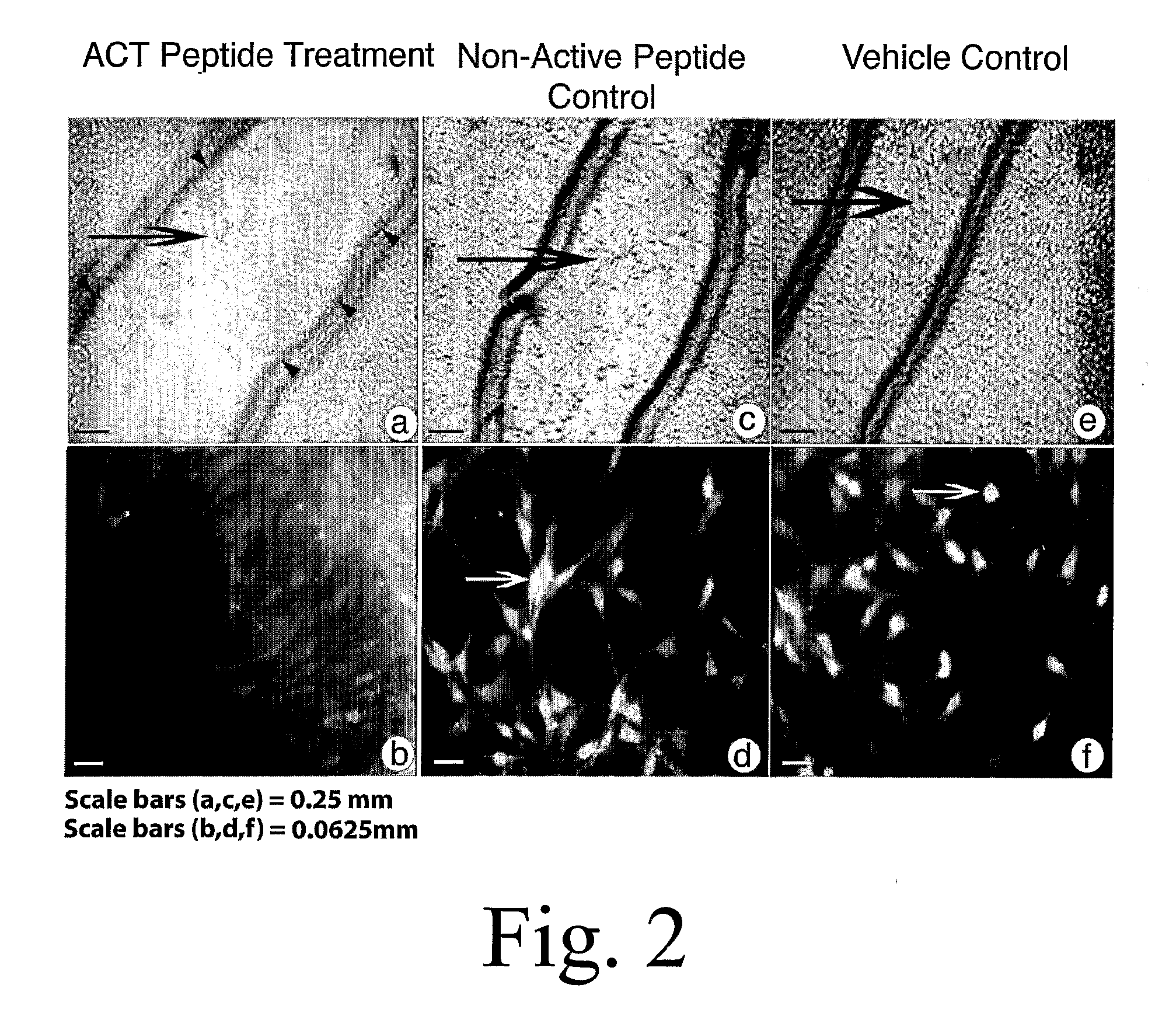
[0155] Myocytes from neonatal rat hearts were grown until forming a near-confluent monolayer on a tissue culture dish according to standard protocols. The cultures were subsequently allowed to culture for a further 5 days culture medium comprising 30 μM ACT 1 peptide (SEQ ID NO:2), 30 μM non-active control peptide (SEQ ID NO:55), or phosphate buffered saline (PBS) containing no ACT peptide or control peptide. The non-active control peptide comprises a polypeptide with a carboxy terminus in which the ACT peptide sequence has been reversed. The amino terminus of ACT and control peptides are both biotinylated, enabling detection (i.e., assay) of the peptides in the cell cytoplasm using standard microscopic or biochemical methods based on high affinity streptavidin binding to biotin.
[0156] Culture media with added peptides or vehicle control was changed every 24 hours during the experiment. FIG. 1a indicates that ACT peptide greatly increased the extent of Cx43 ...
example 2
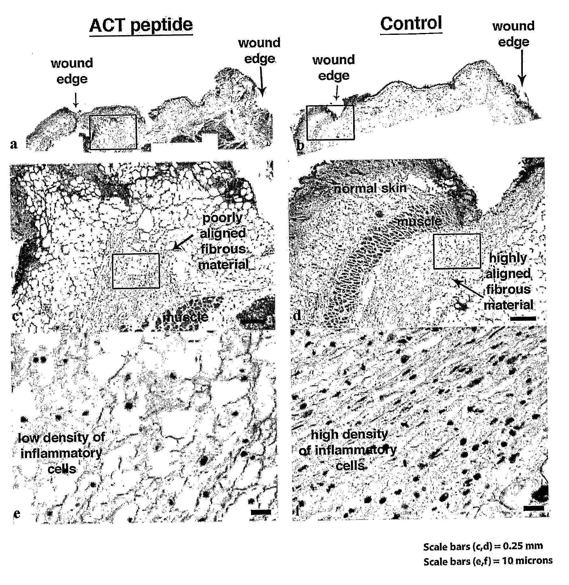
[0160] Neonatal mouse pups were desensitized using hypothermia. A 4 mm long incisional skin injury was made using a scalpel through the entire thickness of the skin (down to the level of the underlying muscle) in the dorsal mid line between the shoulder blades. 30 μl of a solution of 20% pluronic (F-127) gel containing either no (control) or dissolved ACT 1 peptide (SEQ ID NO:2) at a concentration of 60 μM was then applied to the incisional injuries. Pluronic gel has mild surfactant properties that may aid in the uniform dispersion of the ACT peptide in micelles. More importantly, 20% pluronic gel stays liquid at temperatures below 15° C., but polymerizes at body temperature (37° C.). This property of pluronic gel probably aided in the controlled release of peptide into the tissue at the site of incisional injury, protecting the peptide from break-down in the protease-rich environment of the wound and also enabling active concentrations of the peptide to mainta...
example 3
Treatment of Acute Spinal Cord Injury
[0168] Subjects with acute spinal cord injuries represent a seriously problematic group for whom even a small neurological recovery of function can have a major influence on their subsequent independence. In one example, a subject with acute spinal cord injury receives a bolus infusion of a 0.02% to 0.1% solution of ACT peptide (e.g., SEQ ID NO:1) over 15 min within 8 h directly into the site of acute spinal cord injury, followed 45 min later by an infusion of 0.01% solution of ACT peptide for a subsequent 23 to 48 hours. In another example, ACT peptide is used to coat slow release nanoparticles loaded within 8 h directly into the site of acute spinal cord injury or tissue engineered bioscaffolds designed to promote neural reconnection across the zone of acute spinal cord lesion. Improvement in function are assessed by a doctor at intervals (e.g., 6, 12, 26 and 52 weeks) following treatment by neurological outcome tests including assessments des...
PUM
 Abstract
Description
Claims
Application Information
 - R&D
- Intellectual Property
- Life Sciences
- Materials
- Tech Scout
- Unparalleled Data Quality
- Higher Quality Content
- 60% Fewer Hallucinations
Browse by: Latest US Patents, China's latest patents, Technical Efficacy Thesaurus, Application Domain, Technology Topic, Popular Technical Reports.
© 2025 PatSnap. All rights reserved.Legal|Privacy policy|Modern Slavery Act Transparency Statement|Sitemap|About US| Contact US: help@patsnap.com


