
Method for calculation radiation doses from acquired image data

a radiation dose and image data technology, applied in the field of radiation dose determination, can solve the problems of limiting the quality of a reconstructed image, the inability to accurately calculate the dose of radiation from the acquired image, and the inability to accurately use monte carlo methods too computationally expensive for effective clinical us

Inactive Publication Date: 2008-04-17
FAILLA GREGORY A +3
View PDF12 Cites 55 Cited by
  • Summary
  • Abstract
  • Description
  • Claims
  • Application Information

AI Technical Summary

Benefits of technology

[0009] One method embodiment of the present invention is a process for using deterministic methods to calculate dose distributions resulting from radiotherapy treatments, diagnostic imaging, or industrial sterilization, and for calculating scatter corrections used for image reconstruction. One embodiment of the present invention provides a means for constructing a deterministic computational grid from an acquired 3-D image representation of an anatomical region, transporting an external radiation source into the anatomical region, calculating the scattered radiation field in the anatomical region, and calculating the dose field in the anatomical region. Another method embodiment of the present invention includes a process which can enable dose responses in an anatomical region to be calculated, prior to treatment planning, independently of treatment parameters, enabling dose fields to be rapidly reconstructed during treatment plan optimization. In another method embodiment of the present invention, a process to compute the scattered radiation reaching detectors external to the anatomical region is provided. In another method embodiment of the present invention, a process for calculating the radiation field exiting the field shaping components of a radiotherapy treatment head is provided.

Problems solved by technology

For industrial and medical imaging, scattered radiation can substantially limit the quality of a reconstructed image.
The physical models that describe radiation transport through anatomical structures are complex, and accurate methods such as Monte Carlo can be too computationally expensive for effective clinical use.
As a result, most clinically employed approaches are based on simplifications which limit their accuracy and / or scope of applicability.
For imaging, a reduced reconstructed image quality may result.
While Monte Carlo methods are recognized as highly accurate, simulations are time consuming, limiting their effectiveness for clinical applications.
However, the use of deterministic solvers to radiotherapy and imaging applications has been limited to research in radiotherapy delivery modes such as neutron capture therapy and brachytherapy.
This can be attributed to several factors, including methodic limitations in transporting highly collimated radiation sources, and the computational overhead associated with solving equations containing a large number of phase-space variables.
Due to their relative computational efficiency, PBC approaches are widely used in radiotherapy, even though their accuracy is limited, especially in the presence of narrow beams or material heterogeneities.

Method used


Image

  • Method for calculation radiation doses from acquired image data
  • Method for calculation radiation doses from acquired image data
  • Method for calculation radiation doses from acquired image data

Examples


Embodiment Construction

1. Dose Calculations for External Photon Beam Radiotherapy

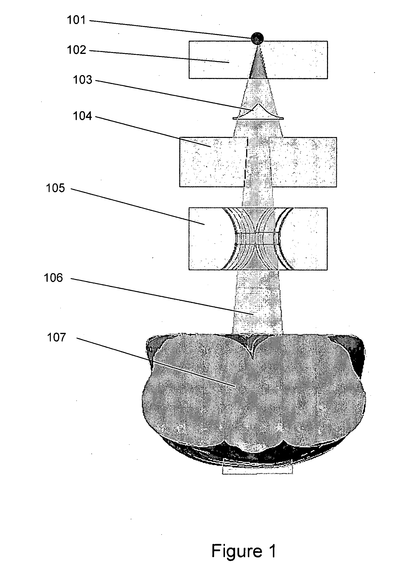
[0056] External photon beam radiotherapy encompasses a variety of delivery techniques, including, but not limited to, 3D-CRT, IMRT, and SRS. FIG. 1 shows a photon radiotherapy beam passing through field shaping components and into an anatomical region. In external photon beam radiotherapy, a photon source 101 may be produced through a number of methods, such as an electron beam impinging on a target in a linear accelerator. This photon source may then be collimated through field-shaping devices, such as the primary collimator 102, flattening filter 103, blocks 104, and multi-leaf collimators 105 to create a beam 106 having a desired spatial distribution that is delivered to an anatomical region 107. The radiation beam may be delivered through a rotating gantry, which may move to multiple positions in the course of a single treatment. By delivering beams from multiple locations, each converging on the treatment region, the h...


PUM


Abstract

Various embodiments of the present invention provide processes for applying deterministic radiation transport solution methods for calculating doses and predicting scatter in radiotherapy and imaging applications. One method embodiment of the present invention is a process for using deterministic methods to calculate dose distributions resulting from radiotherapy treatments, diagnostic imaging, or industrial sterilization, and for calculating image scatter for the purposes of image reconstruction. In one embodiment of the present invention, a method provides a means for transport of external radiation sources through field-shaping devices. In another embodiment of the present invention, a method includes a process for calculating the dose response at selected points and volumes prior to radiotherapy treatment planning.

Description

CROSS-REFERENCE TO RELATED APPLICATIONS [0001] This application is a continuation-in-part of application Ser. No. 11 / 499,862, filed Aug. 3, 2006, which is a continuation-in-part of application Ser. No. 11 / 273,596, filed Nov. 14, 2005, which is a continuation-in-part of application Ser. No. 10 / 910,239, filed Aug. 2, 2004, which is a continuation-in-part of application Ser. No. 10 / 801,506, filed Mar. 15, 2004, which claims the benefit of provisional Application Nos. 60 / 454,768, filed Mar. 14, 2003; 60 / 491,135, filed Jul. 30, 2003; and 60 / 505,643, filed Sep. 24, 2003.TECHNICAL FIELD [0002] The present invention is related to radiation-dose determination and, in particular, computational methods and systems for calculating radiation doses delivered to anatomical tissues and structures from radiotherapy treatments, sterilization processes, or imaging modalities, and for the prediction of scattered radiation related to image reconstruction, for medical and industrial imaging applications....

Claims


Application Information

Patent Timeline
Patent Type & Authority Applications(United States)
IPC IPC(8): G06F17/10G16H20/40
CPCA61N5/1031G06F19/3481A61N2005/1034G16H20/40
Inventor FAILLA, GREGORY A.MCGHEE, JOHN M.WAREING, TODD A.BARNETT, DOUGLAS A.
Owner FAILLA GREGORY A
Learn More