
Biodegradable polymer for marking tissue and sealing tracts

a biodegradable, tissue marker technology, applied in the field of biodegradable tissue marker and sealant, can solve the problems of not knowing how to use tissue marker or sealant as a drug delivery means, and unable to guarantee the migration of tissue markers, so as to improve the modulus of elasticity, slow down the release of drugs, and increase flexibility

Inactive Publication Date: 2008-04-17
BIOPSY SCI LLC
View PDF9 Cites 48 Cited by
  • Summary
  • Abstract
  • Description
  • Claims
  • Application Information

AI Technical Summary

Benefits of technology

[0013] Accordingly, what is needed is a combination tissue marker and sealant that can be formed into many different shapes and sizes, depending upon the application, that can degrade at different rates, depending upon the application, that can expand in response to moisture or other bodily fluids at different rates, depending upon the application, and that does not require in-situ curing.
[0057] Another object is to provide a hydrophobic coating that extends the degradation rate by shielding the substrate from moisture or bodily fluids for a predetermined time.

Problems solved by technology

Anchoring the tissue marker against migration was problematic.
Moreover, although the tissue marker included a contrast solution to facilitate its imaging under X-rays, it was unknown how to use the tissue marker or the sealant as a drug delivery means.
Nor was it known how polymers having utility as tissue markers could be formulated to achieve differing degradation rates.
More particularly, it was not known how to make different polymers to contain contrast for different amounts of time, such as one month to six months or more by using two different polymers.
Nor was it known how to formulate a marker polymer with a therapeutic drug or other pharmaceutical agent so that the degradation of the marker would gradually release the drug or agent to a site.
Nor was it known how the polymers could be formulated to exhibit differing expansion rates when exposed to water or other liquid fluids.
Tissue markers are hard and have little compatibility with surrounding tissue.
One challenge with gene therapy treatments concerns the prevention of immunorejection of genetically modified cells after intracranial implantation.
A further challenge is to achieve efficient gene transfer, as well as prolonged gene expression within the relevant cells.

Method used


Image

  • Biodegradable polymer for marking tissue and sealing tracts
  • Biodegradable polymer for marking tissue and sealing tracts
  • Biodegradable polymer for marking tissue and sealing tracts

Examples


Embodiment Construction

[0095] How to achieve non-covalent bonding of ionic and non-ionic contrast agent with polymers such as PGA / PLA / PCLUPDO will now be described. Different techniques are employed to accomplish similar results in connection with covalent bonding.

[0096] The starting materials employed in this invention for synthesizing the novel tissue marker include poly(DL-lactide), inherent viscosity (IV) of 0.63 dL / g (where the solvent is CHCl3 and the concentration is approximately 0.5 g / dL at 30° C.), 50 / 50 poly(DL-lactide-co-glycolide, IV of 0.17 dL / g (hexafluoroisopropanol, concentration ˜0.5 g / dL at 30° C.) and 75 / 25 poly(DL-lactide-co-glycolides), having IVs of 0.44 dL / g (CHCl3, concentration ˜0.5 g / dL at 30° C.) and 0.69 dL / g (CHCl3, concentration ˜0.5 g / dL at 30° C.). These materials are commercially available from Birmingham Polymers, Inc., of Birmingham, Ala.

[0097] Further starting materials for synthesis of the novel tissue marker include poly(DL-lactide), IV of 1.6 dL / g (CHCl3, concentr...


PUM

PropertyMeasurementUnit
timeaaaaaaaaaa
timeaaaaaaaaaa
concentrationaaaaaaaaaa

Abstract

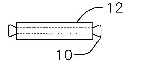
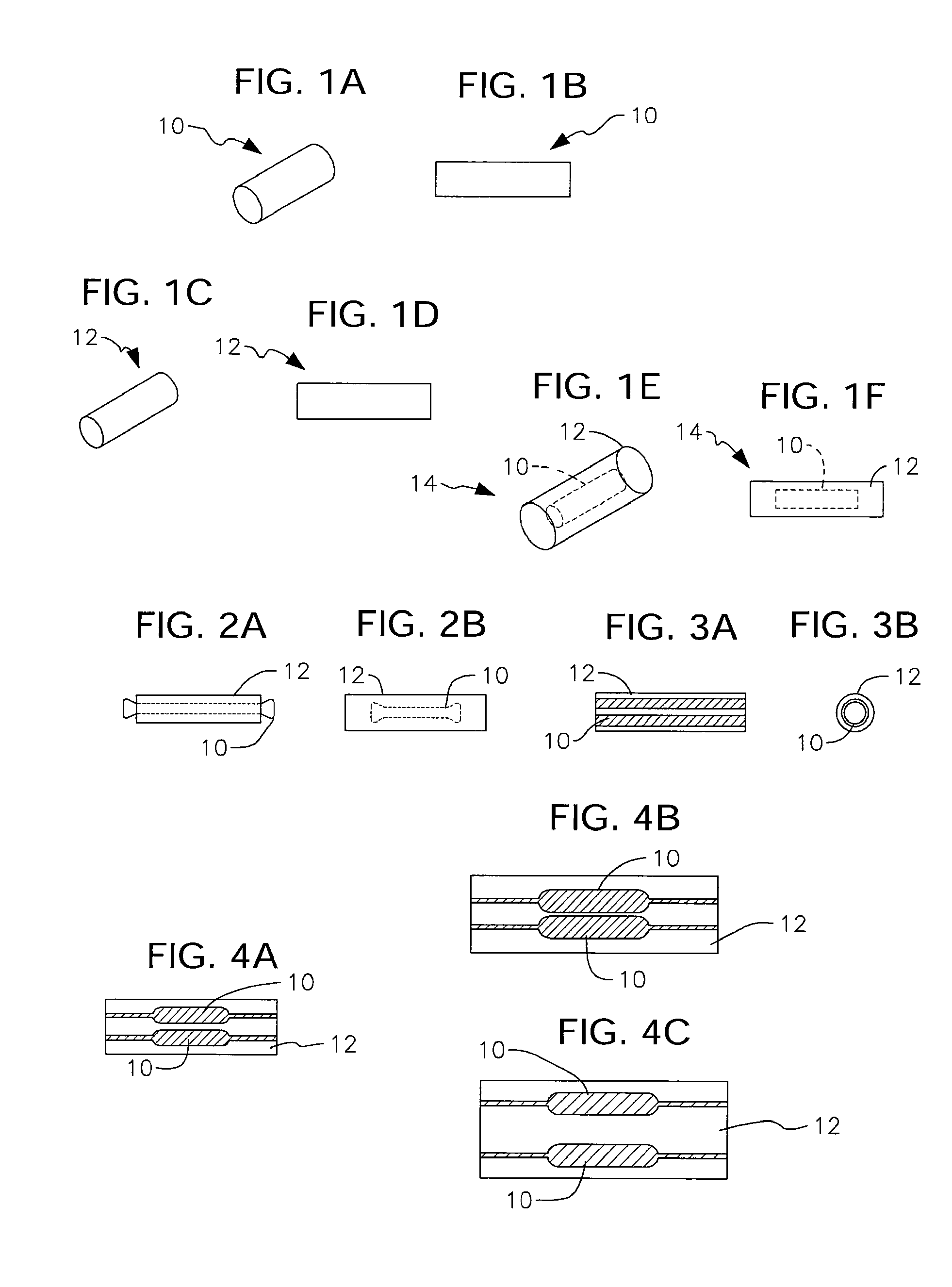
A tissue marker formed of a biodegradable polymer having drug-delivery capabilities is combined with a sealant that encapsulates the tissue marker and which serves to help anchor the tissue marker against migration. The sealant is delivered to a site in dehydrated form and moisture inherent in tissue at the site expands the sealant. The expanded sealant is formed of a hydrogel and is therefore more compatible to the surrounding tissue than the material of the tissue marker. The sealant and the tissue marker are both bioabsorbed over time.

Description

CROSS-REFERENCE TO RELATED DISCLOSURES [0001] This disclosure is a divisional application claiming the benefit of the filing date of pending U.S. patent application entitled: “Biodegradable Polymer for Marking Tissue and Sealing Tracts,” by the same inventors, filed on May 3, 2002, bearing Ser. No. 10 / 063,620.BACKGROUND OF THE INVENTION [0002] 1. Field of the Invention [0003] This invention relates, generally, to a biodegradable tissue marker and sealant. More particularly, it relates to a tissue marker formed of a biodegradable polymer having drug-delivery capabilities. It further relates to a sealant that encapsulates the tissue marker and which serves to help anchor the tissue marker against migration. [0004] 2. Description of the Prior Art [0005] U.S. Pat. No. 6,350,244 entitled Bioabsorable Markers For Use In Biopsy Procedures to the present inventor discloses a bioabsorbable marker that is positioned near a lesion or tumor during a biopsy procedure. The marker includes a contr...

Claims


Application Information

Patent Timeline
Patent Type & Authority Applications(United States)
IPC IPC(8): A61B10/02A61B17/00A61B19/00A61L24/00A61L31/18
CPCA61B19/54A61B2017/00004A61B2017/00893A61B2017/00898A61L24/0015A61L24/0031A61L2300/604A61L31/18A61L2300/406A61L2300/41A61L2300/416A61L2300/44A61L24/0042A61B90/39
Inventor FISHER, JOHN
Owner BIOPSY SCI LLC
Learn More