
Systems for heart treatment

a heart and system technology, applied in the field of minimally invasive, mechanical, medical devices, can solve the problems of ventricular dysfunction, inability to perform the simplest everyday tasks of patients, inability to properly and adequately fill or empty blood from the left ventricle with adequate efficiency to meet the metabolic needs of the body, etc., to facilitate positive or reverse remodeling, improve cardiac output and efficiency, and limit further degeneration of chf

Inactive Publication Date: 2008-04-03
BAY INNOVATION GROUP
View PDF6 Cites 17 Cited by
  • Summary
  • Abstract
  • Description
  • Claims
  • Application Information

AI Technical Summary

Benefits of technology

[0019] Devices and methods according to the present invention not only offer an approach to limit further degeneration of CHF, but variations of the invention can also actively and / or passively facilitate positive or reverse remodeling (i.e., to provide a mild compressive force against the dilated ventricle in synchrony with the pace established by the A-V node) to induce pulsatile contraction of these structures to facilitate improved cardiac output and efficiency. As such, the subject devices and methods provide a potential, palliative or therapeutic response to the referenced disease state.
[0020] Variations or embodiments of the present invention provide cardiac support structures that offer structural rigidity and resistance to overdilation of the cardiac muscle fiber while maintaining an ideal, efficient ventricular shape, orientation of these support structures in specific anatomical positions similar to and in order to restore the helically would native myocardial fiber locations, and application of an energy source to provide active contraction of the myocardium in synchrony with metabolic and functional needs established by the pacemaker driving the electrical activity within the heart.
[0021] A benefit of these cardiac support structures is that they may work in concert to simultaneously provide reinforcement against myocardial stretch (or infarct expansion) and provide an active, positive inotropic during systole. Such devices and associated methods provide dynamic support or reinforcement. Further, they are active throughout the cardiac cycle—unlike previous device-based approaches that solely attempt to passively reduce the stress in the heart wall during diastole. Diastolic compliance can also be regulated or controlled with structures according to the present invention.
[0022] Though not necessarily the case, the cardiac support structures of the invention are typically implanted / deployed using a minimally invasive surgical approach. In practice, the subject structures can be placed via a sub-xiphoid approach which allows sufficient exposure and visualization of the heart using standard minimally invasive means to facilitate placement and anchoring of the support structure(s) at targets zones about the heart.

Problems solved by technology

While the exact etiology of the syndrome that causes heart failure is not fully understood, the primary cause of CHF is the inability of the heart to properly and adequately fill or empty blood from the left ventricle (i.e., left ventricular dysfunction) with adequate efficiency to meet the metabolic needs of the body.
These include neuro-hormonal stimulation, endothelial dysfunction, vasoconstriction, and renal sodium retention all of which can cause dyspnea, fatigue and edema rendering patients unable to perform the simplest everyday tasks.
Even with novel pharmacological, surgical and device-based therapies, symptoms can be alleviated, but the quality of life remains significantly impaired and the associated morbidity and mortality of the disease is exceptionally high.
Decreases in systolic contraction can lead to cardiomyopathy, which further exacerbates the localized, ischemia damaged tissue or AMI insult into a global impairment leading to episodes of arrhythmia, progressive pump failure and death.
Analogous to aneurysms in diseased hearts accompanying abnormally thin and weak myocardial tissue, ischemia-damaged and / or infarct damaged heart muscle tissue results in progressive softening or degeneration of cardiac tissue.
These ischemic and infarcted zones of the heart muscle wall have limited, if not complete loss of tissue contractile functionality and overall physical integrity.
With this enlargement, the heart's burden is increased to pump more blood with each pump cycle.
With this bulging, the heart's natural contraction mechanism is dissipated into and attenuated resulting in a marked and progressing decrease in cardiac output.
The normal mitral valve is a complex structure; consisting of leaflets, annulus, chordae tendonae, and papillary muscles and any damage or impairment in function of any of these key components can render a valve structure incompetent.
Impairment of valve function, due to independent factors (i.e., a concomitant valve pathology) or dependent factors (i.e., valve dilation related to dilated cardiomyopathy or mitral regurgitation due to atrial enlargement), can result in valvular insufficiency further exacerbating the degenerative CHF cycle.
Various pharmacological and surgical methods have been applied both with palliative and therapeutic outcome goals, however there still remains no cure for the condition.
These kinds of vasodilators relax both arterial and venous smooth muscle, thereby reducing the resistance to left ventricular ejection.
The procedure is known to provide some symptomatic improvement, but is controversial with regard to its ability to enable active improvement of cardiac performance.
In spite of the positive outcome on relieving some of the symptoms, the procedure is highly invasive, requiring access to the heart via a sternotomy, expensive, complex and of unknown durability (due to the muscle wrap blood flow requirements and fibrosis issues).
Besides being a highly invasive, traumatic and costly procedure, the actual volume reduction results in a reduction in valve competence and elicits the associated regurgitation.
In dilated hearts resultant of the heart failure cascade the ventricle assumes a more spherical shape and this spiral architecture and hence the associated contractile efficiency is lost.
While being generally successful and routine in surgical practice today, these procedures are also costly, highly invasive, and are still have significant associated morbidity and mortality.
However, the use of such devices is still limited by high costs and a lack of substantial, clinical evidence warranting their use.
As such, these types of approaches require unnecessary positioning of the devices over healthy (non local, undamaged) areas or zones of the heart affecting the entire organ when the primary treatment is usually focused is on the left ventricle or the mitral valve annulus.
This non-localized treatment can elicit iatrogenic conditions such as undesired valvular dysfunction or constrictive physiology due to over restriction of the heart by such restraints.
The clinical efficacy of this approach while appealing is unknown at this time.
Transplants represent a massive challenge with donor hearts generally in short supply and with the transplant surgery itself presenting a high risk, traumatic and costly procedure.
In spite of this, transplants present a valuable, albeit limited, upside increasing life expectancy of end stage congestive heart failure patient from less than one year up to a potential five years.
It is evident that there is currently no ideal treatment among the various surgical, pharmacological, and device based approaches to treat the multiple cardiac and non-cardiac factors implicated with the syndrome of CHF.

Method used


Image

  • Systems for heart treatment
  • Systems for heart treatment
  • Systems for heart treatment

Examples


embodiment 84

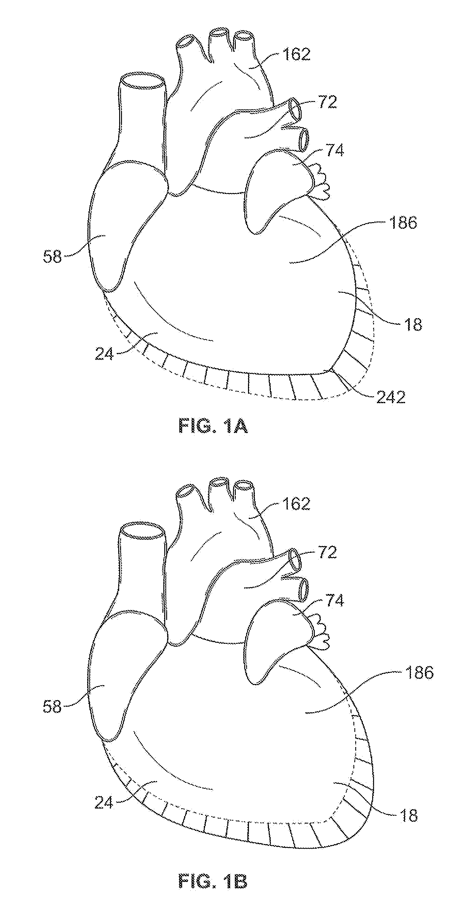
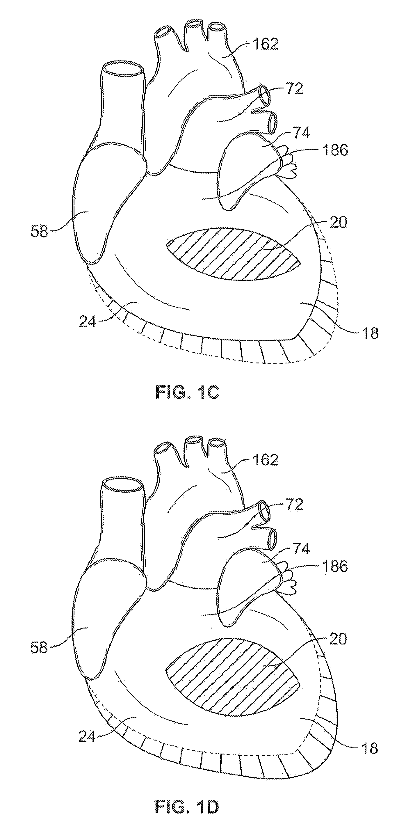
[0053]FIGS. 3A to 3H show a cardiac support structure embodiment 84 that incorporates anchors 32 separately deployable from the tensile members. It should be noted, however, that the anchors can be advantageously be integrated and / or interconnected with the tensile members. FIG. 3C shows an anchor 32 that can be inserted through myocardium and incorporates notches 122 that fit in mating openings 142 of the tensile members to secure it firmly in position for chronic use throughout the necessary cyclic life of the device. The tensile members shown in FIGS. 3A and 3B incorporate a spring component capable (if desired) of a pre-defined extension and contraction such that the tensile member 84 can be expanded during positioning and apply a compressive force against the heart to continuously urge and maintain the heart into the preferred helical orientation or any other orientation as defined by the operator during the implant procedure.

[0054] Tensile members 84 may comprise a tubing of r...

embodiment 4

[0057]FIG. 5B shows cardiac support structures 4 as shown in FIG. 5A deployed through the myocardium of heart 186. In the cardiac support structure embodiment 4 in FIG. 5A, a single tensile member 84 is shown with spaced apart tissue penetration ends 120 on one side and a connecting spring on the opposite side 126. Upon insertion of the tissue penetrating ends 120 of the tensile members 84, a cardiac support structure anchor 124 can be placed over these ends to secure the member to the tissue surface or the ends of the tensile member can be tied to produce an axially-oriented tightening of the support structure 4. Still further, one free end of the tensile member can be subsequently inserted through myocardium at a spaced apart location to produce a three-dimensional, cinching effect.

[0058] According to one aspect of the invention, multiple cardiac support structures are secured to the heart tissue to produce a helical pattern as shown in FIG. 5B, thereby maintaining or repositionin...


PUM


Abstract

Devices and methods for treating degenerative, congestive heart disease and related dysfunction are described. Passive and active cardiac support structures mitigate changes in ventricular structure (i.e., remodeling) and deterioration of global left ventricular performance related to tissue damage precipitating from ischemia, acute myocardial infarction (AMI) or other abnormalities. Cardiac efficiency is improved by providing reinforcement that restores or maintains an elliptical ventricular shape and mimics the position and positive inotropic effects of helical wound myofibrils to provide active contraction of the ventricle in synchrony with the metabolically required cardiac pace or output. In addition, the cardiac support structures compensate or provide therapeutic treatment for congestive heart failure and / or reverse the remodeling that produces an enlarged heart. The structures may be implanted in target heart regions using less invasive surgical techniques, such as those involving port access or small incisions into the thoracic cavity.

Description

CROSS REFERENCE TO RELATED APPLICATIONS [0001] This application claims the benefit of co-pending Provisional Patent Application Ser. No. 60 / 519,915, filed Nov. 14, 2004 and entitled, “Minimally Invasive Systems for Heart Constraint and Reshaping with Passive or Active Contraction” which is incorporated by reference herein in its entirety.FIELD OF THE INVENTION [0002] The present invention relates generally to minimally invasive, mechanical, medical devices for treating or preventing congestive heart failure and related or concomitant vascular dysfunction. More specifically, the invention relates to cardiac support structures that mitigate changes in the ventricular and / or atrial structure and geometry and deterioration of global left and right ventricular and atrial performance related to tissue damage from myocardial ischemia, acute myocardial infarction (AMI), valve related disease or dysfunction, vascular related dysfunction, or other instigators of deterioration of cardiac outpu...

Claims


Application Information

Patent Timeline
Patent Type & Authority Applications(United States)
IPC IPC(8): A61N1/362A61N1/05A61F2/00A61F2/24
CPCA61F2002/249A61F2/2481
Inventor PAI, SURESHDOMINGO, NICANOR
Owner BAY INNOVATION GROUP
Learn More