Arm brace for sonographers

a technology for sonographers and arm braces, applied in the direction of tomography, diagnostic recording/measuring, rod connection, etc., can solve the problems of work-related injuries, difficult gripping of the probe, epidemic among medical sonographers, etc., and achieve the effect of reducing or eliminating prolonged and repeated “pinch and push” activity

Active Publication Date: 2008-02-28
WISCONSIN ALUMNI RES FOUND
View PDF12 Cites 8 Cited by
  • Summary
  • Abstract
  • Description
  • Claims
  • Application Information

AI Technical Summary

Benefits of technology

[0007]The present invention provides a support that transfers force directly between the ultrasound probe and the sonographer's forearm and/or upper arm and shoulder, bypassing the sonographer's wrist. In a preferred embodiment, the ultrasound probe is flexibly connected to an arm bra...

Problems solved by technology

The probe body is normally constructed of a hard, seamless polymer shell protecting the probe electronics and allowing for easy cleaning of the probe, but making the probe difficult to grip.
This problem can be exacerbated by the acoustic coupling gel normally used to increase acoustic co...

Method used


Image

  • Arm brace for sonographers
  • Arm brace for sonographers
  • Arm brace for sonographers

Examples


Embodiment Construction

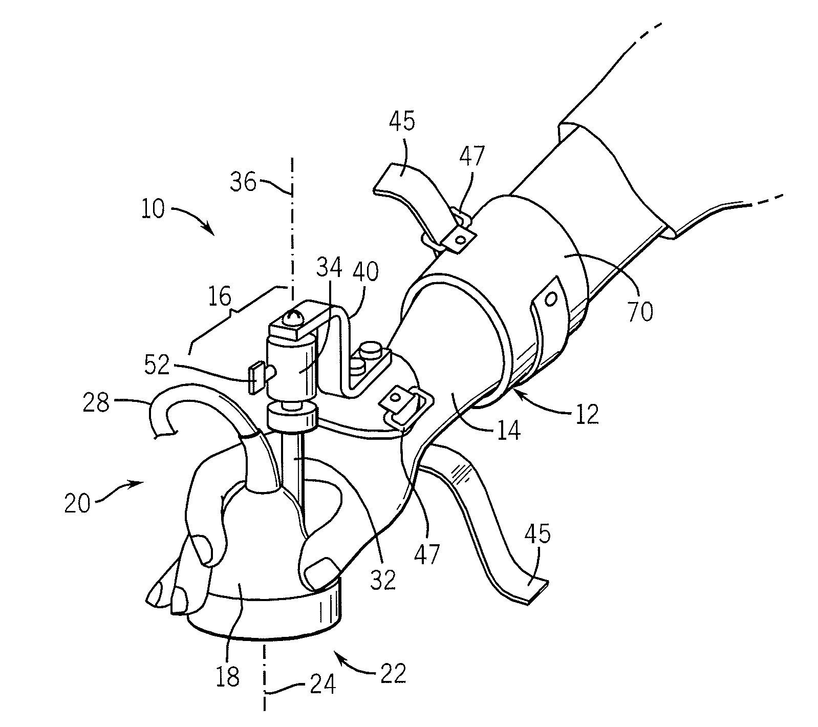
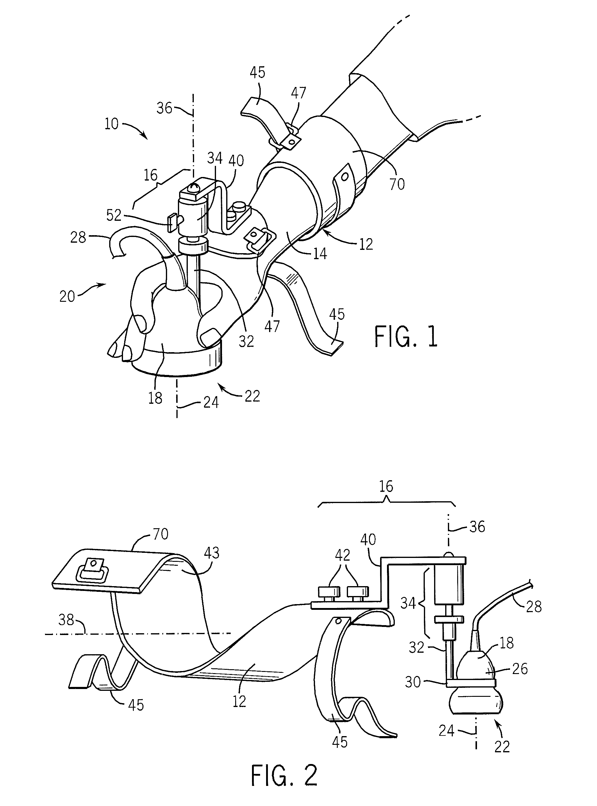
[0040]Referring now to FIG. 1, an ultrasound probe support 10 of the present invention may include generally an arm brace 12 for attachment to the forearm 14 of a sonographer. A distal end of the arm brace 12 supports a coupling 16 that communicates between the arm brace 12 and an ultrasound probe 18 that may be held by the sonographer's hand 20.

[0041]Referring now also to FIG. 2, the ultrasound probe 18 will generally have an axially extending probe body 26 terminating at one end at a probe face 22 from which ultrasonic signals are emitted and echo signals are received along an ultrasound axis 24. A signal cord 28 may extend from the opposite end of the probe body 26, the signal cord attaching the ultrasound probe 18 to an ultrasound machine (not shown) which processes received ultrasonic data to produce images as is understood in the art.

[0042]The ultrasound probe 18 is connected to one end of the coupling 16 by means of a grip 30 removably connected to the body 26 of the ultrasou...


PUM


Abstract

A support for sonographers provides a path of force transmission from an ultrasound probe to the sonographer's upper arm bypassing the wrist, and thus reducing wrist injuries that can come from the need to tightly grip and restrain an ultrasound probe for long periods of time.

Description

CROSS-REFERENCE TO RELATED APPLICATIONS[0001]The present application claims the benefit of U.S. Provisional Patent Application Ser. No. 60 / 840,663 filed on Aug. 28, 2006.STATEMENT REGARDING FEDERALLY SPONSORED RESEARCH OR DEVELOPMENTBACKGROUND OF THE INVENTION[0002]The present invention relates to apparatus for supporting ultrasound probes in medical imaging and, in particular, to a brace intended to reduce occupational injuries to sonographers.[0003]Ultrasound medical imaging uses ultrasound sound waves to construct an image of internal body structures. During an ultrasound imaging procedure, an ultrasonic transducer (probe) is pressed against a patient to transmit ultrasound into the patient and to receive echoes that may be analyzed to produce the image. The probe is typically hand held by a sonographer to be easily manipulated to direct the ultrasound to a desired region of interest and / or to sweep the direction of the ultrasound to create a dynamic image of internal body struct...

Claims


Application Information

Patent Timeline
IPC IPC(8): A61B8/14A61F5/00
CPCA61B5/6824A61B8/4281A61F5/0118Y10T403/291Y10T403/32557Y10T403/32606Y10T403/32631Y10T403/32565Y10T403/1666Y10T403/32311Y10T403/32549A61B8/4227
Inventor KLIEWER, MARK A.WALKER, TIMTHOMAS, CHRISTOPHER J.
Owner WISCONSIN ALUMNI RES FOUND
Why Eureka
Try Eureka