
Clip applier and methods of use

a technology which is applied in the field of applicator and clip, can solve the problems of time-consuming and expensive procedures, requiring as much as an hour of physicians or nurses time, and uncomfortable for patients, and achieves the effect of enhancing the hemostasis within the opening

Active Publication Date: 2008-01-03
ABBOTT LAB INC
View PDF101 Cites 321 Cited by
  • Summary
  • Abstract
  • Description
  • Claims
  • Application Information

AI Technical Summary

Benefits of technology

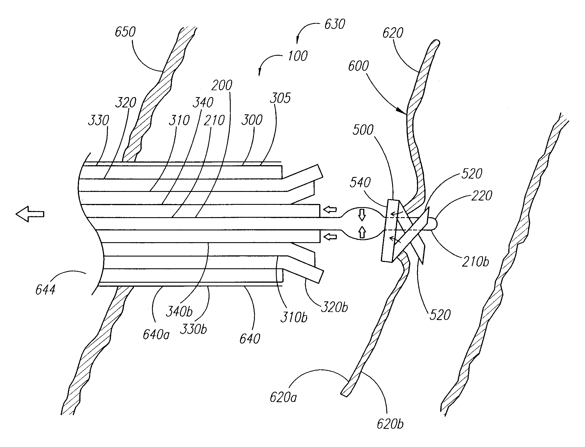
[0011]When properly positioned, the apparatus can be activated to distally deploy the closure element. During deployment, the apparatus can be configured to substantially uniformly expand the closure element beyond a natural cross-section of the closure element such that the closure element, when deployed, can be configured to engage the blood vessel wall and / or tissue. Engaging the blood vessel wall and / or tissue, the closure element can be further configured to return to the natural cross-section. Thereby, the engaged blood vessel wall and / or tissue are drawn substantially closed and / or sealed, such that, for example, hemostasis within the opening can be enhanced.
[0012]The present invention can also accommodate for variations in the size of the physicians hand and grip by selectively reducing the distance between the devices handle portion and a portion of a triggering system usable to deploy the closure element. The triggering system of the apparatus can at least partially move a trigger extension graspable by a physician or clinician as a locator assembly locates the blood vessel wall prior to deploying the closure element. This partial movement reduces the gap between the trigger extension and the handle portion. In this manner, a physician or clinician does not need to stretch uncomfortably to position a thumb or finger on the trigger extension, grasping the handle portion, and maintaining the device in the desired orientation relative to the tissue and / or the puncture site.
[0015]The present invention can also provide a stable base upon which the physician or clinician can move the device or apparatus as the closure element is positioned and deployed. In one configuration, the stable base is formed from the handle portion having two graspable portions: a shaped grasping portion and an elongated grasping portion. The shaped grasping portion can be configured to receive at least a thumb or finger of the physician. A portion of the handle portion can have a curved profile to enable a portion of the handle to fit comfortably within a user's hand while the hand is rested on a patient during the procedure to provide stability during use of the device and function as a based or pivot point for moving the remainder of the device or apparatus.

Problems solved by technology

This procedure, however, may be time consuming and expensive, requiring as much as an hour of a physicians or nurses time.
It is also uncomfortable for the patient, and requires that the patient remain immobilized in the operating room, catheter lab, or holding area.
In addition, a risk of hematoma exists from bleeding before hemostasis occurs.
This device, however, requires that an anchor of the plug be positioned within the vessel, and therefore, may increase the risk of over-advancement of the plug itself into the vessel.

Method used


Image

  • Clip applier and methods of use
  • Clip applier and methods of use
  • Clip applier and methods of use

Examples


Embodiment Construction

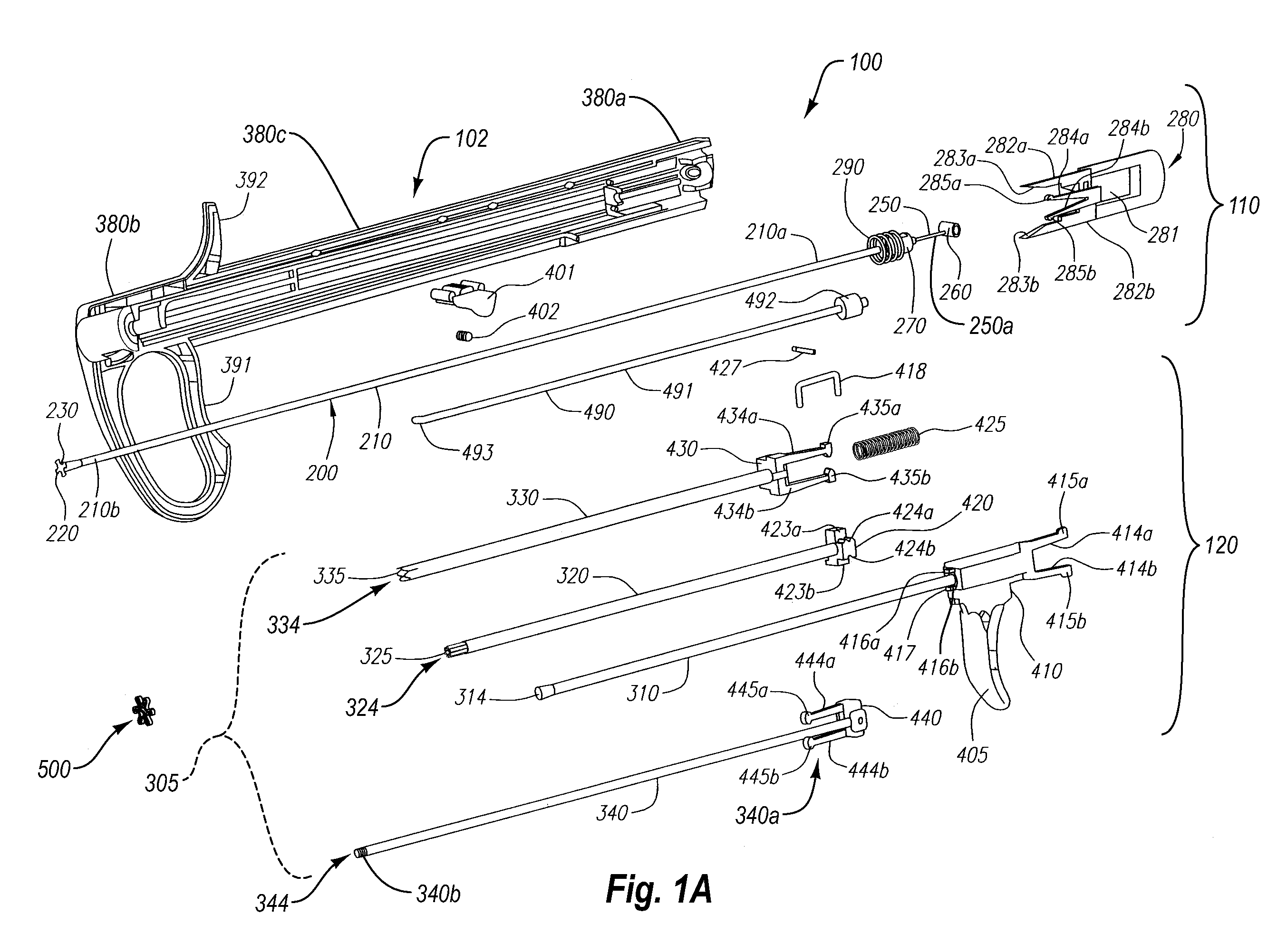
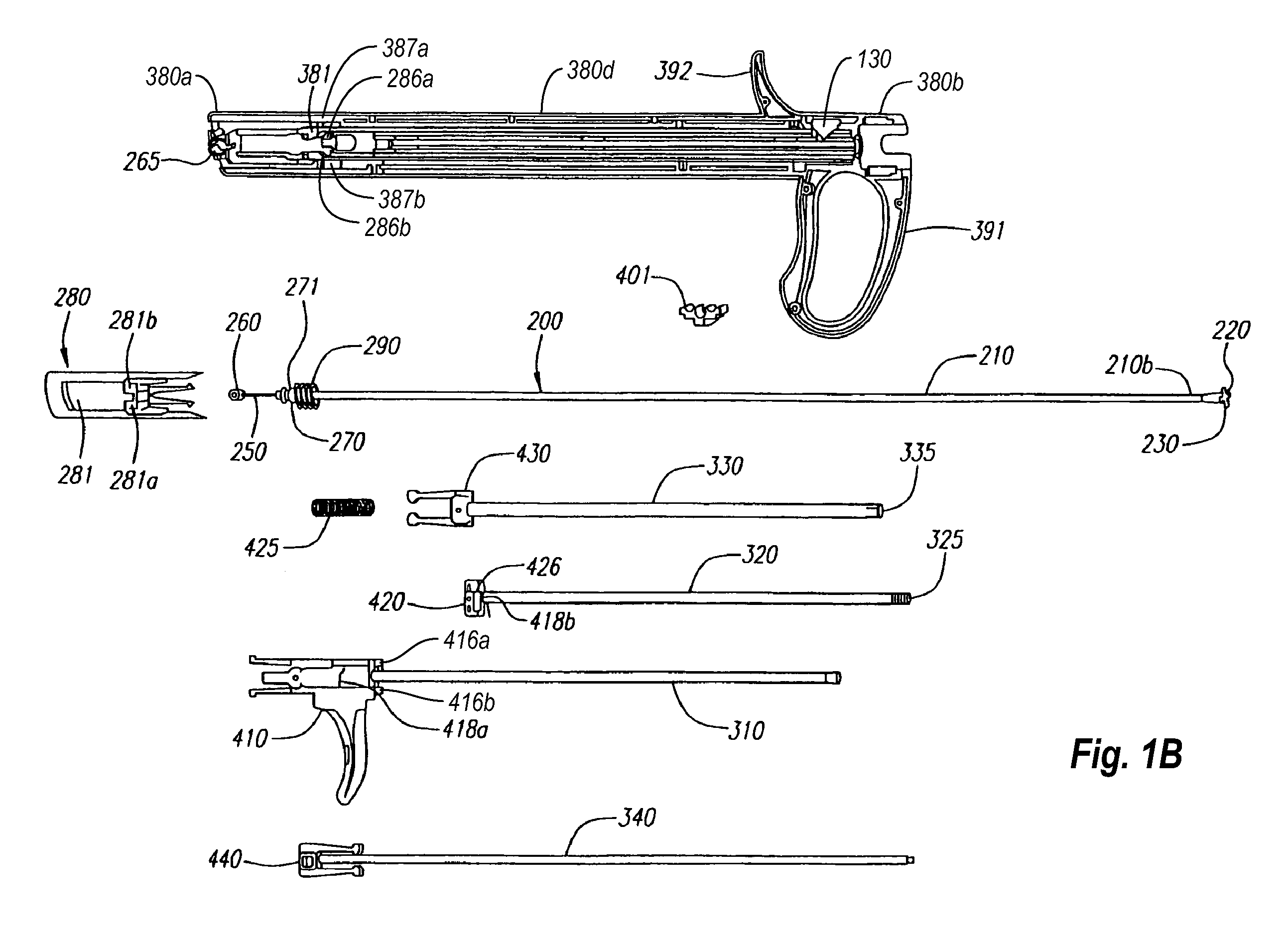
[0042]The embodiments described herein extend to methods, systems, and apparatus for closing and / or sealing openings in a blood vessel or other body lumen formed during a diagnostic or therapeutic procedure. The apparatuses of the present invention are configured to deliver a closure element through tissue and into an opening formed in and / or adjacent to a wall of a blood vessel or other body lumen.

[0043]Since current apparatuses for sealing openings formed in blood vessel walls can snag tissue adjacent to the openings during positioning and may not provide an adequate seal, an apparatus that is configured to prevent inadvertent tissue contact during positioning and to engage tissue adjacent to the opening can prove much more desirable and provide a basis for a wide range of medical applications, such as diagnostic and / or therapeutic procedures involving blood vessels or other body lumens of any size. Further, since current apparatuses for sealing openings formed in blood vessel wal...


PUM


Abstract

An apparatus for placement into an opening formed in a wall of a body lumen to deploy a closure element. The apparatus comprising a housing having a handle portion formed at a distal end, the handle portion configured to be engaged by a user when advancing the housing to deploy the closure element. A locator assembly and a carrier assembly are at least partially received in the housing. The locator assembly can have one or more expansion members that can selectably contact the wall of the body lumen and a proximal end configured to cooperate with a portion of a locator control system operable to expand the one or more expansion members. A triggering system cooperates with the locator assembly and is moveable toward the one or more expansion members of the locator assembly upon the locator control system being operated to expand the one or more expansion members.

Description

CROSS-REFERENCE TO RELATED APPLICATIONS[0001]This application claims priority to U.S. Provisional Patent Application, Ser. No. ______, filed Jul. 1, 2005, which is incorporated herein by reference in its entirety. This application also incorporates by reference U.S. patent application Ser. No. 10 / 356,214 and U.S. patent application Ser. No. 10 / 638,115 in their entireties.FIELD OF THE INVENTION[0002]The present invention relates generally to apparatus and methods for closing and / or sealing openings through tissue, and more particularly to apparatus and methods for delivering a closure element for closing a puncture in a blood vessel or other body lumen formed during a diagnostic or therapeutic procedure.BACKGROUND OF THE INVENTION[0003]Catheterization and interventional procedures, such as angioplasty or stenting, generally are performed by inserting a hollow needle through a patient's skin and tissue into the vascular system. A guide wire may be advanced through the needle and into ...

Claims


Application Information

Patent Timeline
IPC IPC(8): A61B17/10
CPCA61B17/0057A61B17/064A61B17/0644A61B17/068A61B2017/00672A61B17/10A61B17/1227A61B2017/00424A61B17/083
Inventor WALBERG, ERIK K.REYNOLDS, TIMOTHY C.ELLINGWOOD, BRIAN A.JAIR, KAI Y.PANTAGES, ANTHONY
Owner ABBOTT LAB INC
Learn More