
Method and system for automatic identification and orientation of medical images

a technology of automatic identification and orientation of medical images, applied in the field of computer assisted detection (cad), can solve the problems of insufficient system for loading and feeding film-based medical images to the scanner, requiring too much time and effort from the user, and unable to diagnose, so as to achieve time saving and reduce errors

Active Publication Date: 2007-04-05
HOLOGIC INC
View PDF5 Cites 26 Cited by
  • Summary
  • Abstract
  • Description
  • Claims
  • Application Information

AI Technical Summary

Benefits of technology

The present invention provides an automated and computerized method for detecting the order and orientation of a film series using predetermined film identifying marks. This allows a technologist to feed films into the system in any order and orientation while providing an output to a radiologist that is in an order and orientation preferred by the radiologist. Additionally, the invention distinguishes films belonging to one case from those of another case, which is useful when a large number of cases are fed into a digitizer at one time with a bulk loading input feeder. The technical effects of this invention include time savings, reduction of errors, and improved efficiency in processing large volumes of films.

Problems solved by technology

While not a replacement for the experienced radiologist, CAD systems are designed to increase efficiency and reduce error, as a typical radiologist may be required to examine hundreds of medical images per day, which can lead to the possibility of a missed diagnosis due to human error.
With current CAD systems, however, we have found that systems for loading and feeding film-based medical images to the scanner are inadequate in that they tend to require too much time and effort from the user.
Additionally, we have found that currently available systems do not allow the user to simply and conveniently enter case information, monitor the status of cases, and abort or adjust the processing of films being processed.
For example, with currently available systems, errors in film orientation may not be detected until the radiologist views the analyzed image.
Experience has shown, however, that this requirement places some burden on the technologist operating the system, adding to the time required to do the work, and occasionally being a source of error.
For the technologist to align all films in each case in a prescribed manner would be onerous, time consuming, and prone to error.

Method used


Image

  • Method and system for automatic identification and orientation of medical images
  • Method and system for automatic identification and orientation of medical images
  • Method and system for automatic identification and orientation of medical images

Examples


Embodiment Construction

[0027] It has been found that the currently available CAD systems are lacking in their ability to facilitate the loading and feeding of films for processing. Furthermore, it has been found that such systems further lack in their ability to display digitized images to a doctor or radiologist. In particular, it has been found that providing a film feeding mechanism that holds multiple films and automatically feeds the films to the scanner greatly reduces the time and labor required to load and input films into the system. It has been found that a stack film feeder that holds a relatively large number of films makes the inputting of films much more efficient. Furthermore, it has been found that a system that allows for displaying digitized films in various orders and / or orientations provides for a more user-friendly interface.

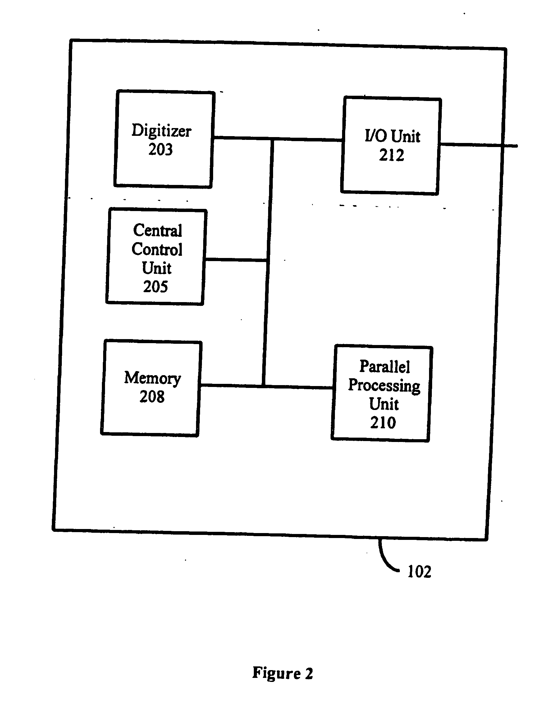
[0028]FIG. 1 shows an outside view of a computer aided detection (CAD) system 100, such as the Image Checker M1000 from R2 Technology, Inc., for assisting in the...


PUM


Abstract

A method and system for computer-aided detection of abnormal lesions in digital mammograms is described, wherein digital films are processed using an automated and computerized method of detecting the order and orientation of a set of films. In one embodiment, anatomic features are used to detect the order, orientation and identification of a film series. In another embodiment of the invention, a technologist feeds films into the system in any order and orientation. After processing, the system provides an output on a display device to a radiologist that is in an order and orientation preferred by the radiologist. In yet another embodiment of the invention, films from one case are distinguished from films of another case. In this manner and through the use of a bulk loader, a large number of films can be stacked together and fed into the system at one time.

Description

FIELD OF THE INVENTION [0001] The present invention relates to the field of Computer-Aided Detection (CAD) of abnormalities found in medical images and to the field of Picture Archiving and Communication Systems (PACS). In particular, the invention relates to a method and system for determining the orientation of digitized medical images in order to enable consistent and understandable communication between a computer-aided detection system and an operator. The invention further relates to a method and system for uniquely identifying and distinguishing images from a particular case. BACKGROUND OF THE INVENTION [0002] Systems for computer-aided detection (“CAD”) assist radiologists in the detection and classification of abnormal lesions in medical images. The purpose of such devices, as described in U.S. Pat. No. 5,815,591 to Roehrig, et. al., entitled “Method and Apparatus for Fast Detection of Spiculated Lesions in Digital Mammograms,” the disclosure of which is hereby incorporated...

Claims


Application Information

Patent Timeline
Patent Type & Authority Applications(United States)
IPC IPC(8): G06K9/00G06T7/00
CPCG06T7/0012G06T2207/30068G06T7/0042G06T7/73
Inventor HARTMAN, KEITHMARSHALL, JULIANSCHNEIDER, ALEXANDERROEHRIG, JIMMY
Owner HOLOGIC INC
Learn More