Modified colony assay

a technology of modified colony and assay, which is applied in the field of hematopoietic cfcassay, can solve the problems of inexperienced laboratory personnel, inability to distinguish between colony types on the basis of differences in morphology or degree of hemoglobinization, and inability to use existing cfc assay formats to obtain information, etc., to promote the development of one colony type, prevent or suppress the development of other colony types, and promote the development of erythroid

Inactive Publication Date: 2007-03-15
STEMCELL TECHNOLOGIES
View PDF2 Cites 4 Cited by
  • Summary
  • Abstract
  • Description
  • Claims
  • Application Information

AI Technical Summary

Benefits of technology

[0019] In one aspect, the invention comprises adding a detection reagent, preferably a fluorescently labeled antibody, that is specific for antigens (preferably a cell-surface antigen) expressed on progenitors or on specific mature cell types to a culture at the start or during culture. Examples of antigens include GPA for erythroid cells, CD45 for white blood cells and CD34 for progenitors. The labeled antibody is present throughout subsequent culture, and binds to colonies while they develop and express the antigen for which the antibody is specific. By periodically inspecting the colonies using an inverted fluorescence microscope, specific colony types can be identified as they start expressing lineage-specific antigens, bind the labeled antibody and become fluorescent. Other colonies, that do not express the antigen remain non fluorescent, while colonies that lose expression of certain antigens during development may be fluorescent at early time points, but lose fluorescence later. The invention thus enables the researcher to distinguish colonies on the basis of antigen expression as well as size and morphology. This facilitates accurate and rapid identification and enumeration of specific colony types at the end of culture, and also enables the researcher to follow the kinetics of the development of different colony types during culture on the basis of changes in antigen expression. This is an improvement over the standard CFC assay method of detecting colonies which uses size and morphology, which is inherently subjective and wherein certain colonies can only be detected at the end of the culture period.
[0024] The invention also comprises modifications to the culture medium and cell preparations used in standard CFC assays to selectively promote the development of one colony type while preventing or suppressing the development of other colony types. In one embodiment, the development of erythroid colonies is promoted and the development of myeloid colonies inhibited by use of serum free medium supplemented with the cytokines SCF and EPO. The absence of serum prevents background colony formation caused by cytokines or other stimulatory compounds present in fetal bovine sera used commonly in culture assays. In another embodiment, background colony formation in the absence of cytokines is prevented or inhibited by using modified cell preparations from which accessory cells, which do not form colonies themselves but can support the growth of specific colony types, are removed by depleting specific cell types, e.g., monocytes, or by enriching progenitor cells.

Problems solved by technology

Inconsistent scoring can result from differences in the criteria for colony identification between groups and inexperience of laboratory personnel (Burger et al., Transfusion (1999); Lumley et al.
Individual colonies can be identified at earlier time points during culture, but distinction between colony types on the basis of differences in morphology or degree of hemoglobinization is not possible as the cells have not been sufficiently matured to be morphologically distinguishable.
Therefore, existing CFC assay formats cannot be used to obtain information about the development of different lineages at various time points during the course of the cultures.
Such approaches can in principle be used to identify different colony types in hematopoietic CFC assays, but available detection methods, e.g., flow cytometry, immunofluorescence microscopy and immunocytochemistry, can only be performed at the end of the culture period as they involve harvesting and breaking up of individual colonies (for flow cytometry), or require drying and chemical fixation of the whole cultures before staining colonies in situ.
Drying down and staining of colonies can be performed for CFC assays done in collagen or agar-based media, but it is technically difficult to preserve colony morphology and prevent nonspecific staining.
This approach cannot be performed in the majority of hematopoietic CFC assays, which are done in methylcellulose-based medium, since colony morphology is completely destroyed.
One problem with the standard CFC assay is background CFC development due to cytokines and other compound in the medium.
Another problem with the standard CFC assay is background CFC formation due to cytokines produced by accessory cells present in the cell preparation.
For these reasons, the neurosphere assay may not provide reliable information on clonal growth properties and frequencies of neural stem cells and progenitors.

Method used


Image

  • Modified colony assay
  • Modified colony assay
  • Modified colony assay

Examples


example 1

Preparation of a Standard Hematopoietic CFC Assay

[0065] Human BM, CB or MPB mononuclear cells were suspended in a methylcellulose-based culture medium containing cytokines (MethoCult™ H4434, StemCell Technologies) (StemCell Technologies Methocult™ Flyer; StemCell Technologies 2003 Catalogue pp. 86-125) at a concentration of between 1×10⁴ and 1×10⁵ cells per 1.1 mL of medium. The culture medium was then dispensed into 35 mm culture dishes using 3 mL syringes with blunt end needles, with 1.1 mL of medium per dish. All cultures were incubated for a total of 14 days at 37° C., 5% CO2, 100% humidity. Colonies were enumerated and classified by bright-field microscopy into the following categories based on size and morphology: CFU-E, BFU-E, CFU-GM, CFU-GEMM (StemCell Technologies Colony Atlas).

example 2

Staining of Erythroid Colonies With Fluorescent Antibody Conjugates

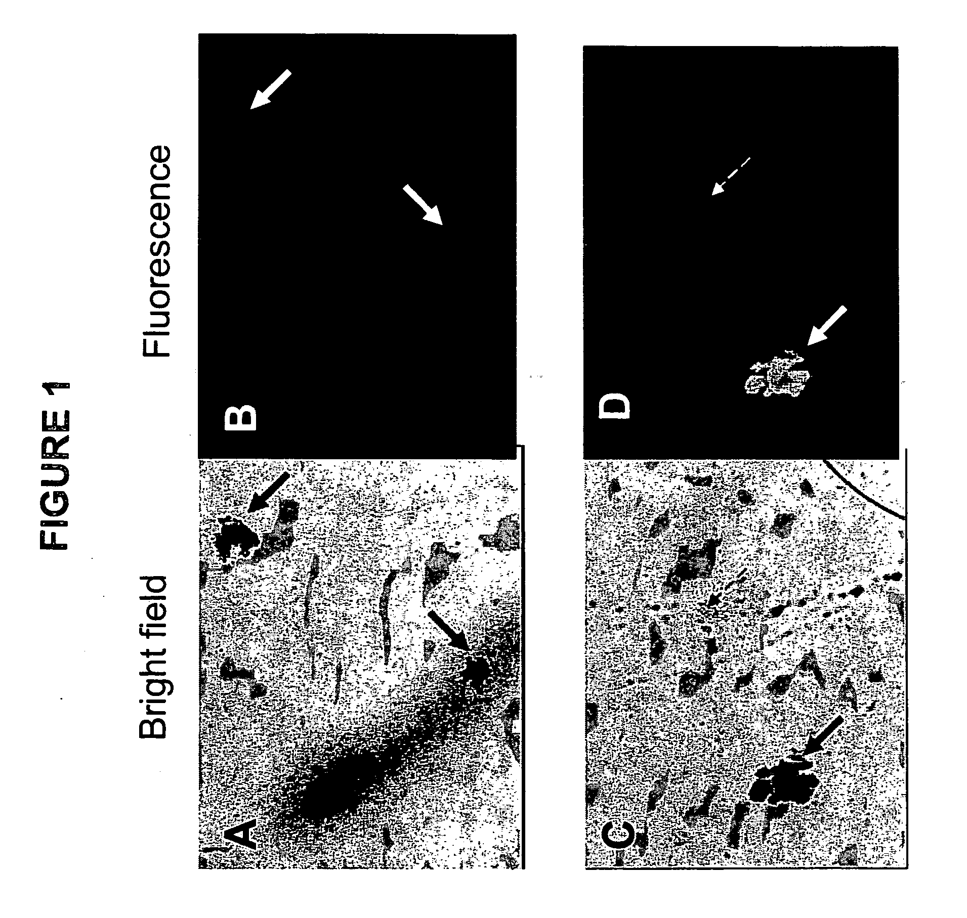
[0066] Standard CFC assays were prepared as described in example 1. At the time of plating, 1 to 10 μg of a FITC-conjugated antibody against Glycophorin-A (anti-GpA, catalogue number 10423, StemCell Technologies) was added per mL of medium. GpA is expressed on erythroid cells and is absent on other cell types, including myeloid cells. As a negative control separate dishes were stained in an identical manner with a FITC-conjugated control antibodies against a marker that is not expressed on eukaryotic cells (IgG2b-FITC, catalog number 557701, Becton-Dickinson, San Jose, Calif.). After 14 days of culture at 37° C. the dishes were inspected using an inverted fluorescence microscope. FIG. 1 shows photographs of section of dishes that were inspected at high magnification using either bright field illumination (FIGS. 1A and 1C) or ultra-violet (UV) illumination to detect fluorescence (FIGS. 1B and 1D). The photographs in ...

example 3

Monitoring of Erythroid Colony Development and Changes in Erythroid Antigen Expression During Culture.

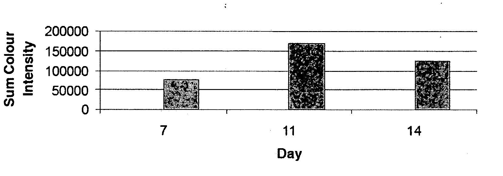
[0067] A CFC-assay was set up as described in Example 2. The culture dishes were inspected daily and the fluorescence of selected colonies were imaged at various time points. All microscope and image capture settings were kept constant. Colonies were analysed by densitometry using AnalySIS software (Soft Imaging System Corp., Riverside, Calif.) and fluorescence intensity was calculated for each time point. FIG. 3 shows that fluorescence of an anti-GPA-FITC stained erythroid colony was detectable as early as day 7, reached maximum intensity at day 11 and declined at the end of the culture period at day 14.


PUM


Abstract

Improved colony forming cell (CFC) assays are described. The improved assay comprises modifications to the standard CFC assay that enable analysis of temporal, real-time changes in antigen expression during colony development without need to fixate or destroy the culture. The improved assay is applicable to hematopoietic CFC assays as well as to CFC assays for other cells, such as neural cells and mammary cells. In one embodiment, the invention comprises adding a detection reagent, most likely a fluorescently labeled antibody, that is specific for antigens (most likely a cell-surface antigen) expressed on progenitors or on specific mature cell types to a culture at the start or during culture. The invention also comprises modifications to the culture medium and cell preparations used in standard CFC assays to selectively promote the development of one colony type while preventing or suppressing the development of other colony types.

Description

[0001] This application claims the benefit under 35 U.S.C.§119(e) of U.S. provisional patent application No. 60 / 715,579 filed Sep. 12, 2005, which is incorporated herein by reference in its entirety.BACKGROUND OF THE INVENTION Hematopoietic CFC-assays [0002] In the adult, hematopoiesis is maintained by the constant regulated production of billions of mature blood cells derived from a small population of relatively quiescent hematopoietic stem cells (HSC) located in the bone marrow. These cells have the potential to undergo self-renewal to maintain their numbers and to produce cells of all the hematopoietic lineages. Proliferation and differentiation of HSC produces a heterogeneous compartment of actively dividing cells called hematopoietic progenitors. With progressive cell divisions, these progenitors have limited self-renewal capacity and become committed to the various blood cell lineages i.e. white blood cells, red blood cells and platelets. [0003] Since their introduction more...

Claims


Application Information

Patent Timeline
IPC IPC(8): G01N33/567
CPCG01N33/56972G01N33/56966
Inventor WOGNUM, ALBERTUS WERNERUSLOUIS, SHARON A.DOSSANTOS, GARY PETERTHOMAS, TERRY E.
Owner STEMCELL TECHNOLOGIES
Why Eureka
Try Eureka